array:24 [
  "pii" => "S2531043720302671"
  "issn" => "25310437"
  "doi" => "10.1016/j.pulmoe.2020.12.007"
  "estado" => "S300"
  "fechaPublicacion" => "2021-07-01"
  "aid" => "1579"
  "copyright" => "Sociedade Portuguesa de Pneumologia"
  "copyrightAnyo" => "2021"
  "documento" => "simple-article"
  "crossmark" => 1
  "licencia" => "http://creativecommons.org/licenses/by-nc-nd/4.0/"
  "subdocumento" => "cor"
  "cita" => "Pulmonol. 2021;27:371-3"
  "abierto" => array:3 [
    "ES" => true
    "ES2" => true
    "LATM" => true
  ]
  "gratuito" => true
  "lecturas" => array:1 [
    "total" => 0
  ]
  "itemSiguiente" => array:19 [
    "pii" => "S2531043721000015"
    "issn" => "25310437"
    "doi" => "10.1016/j.pulmoe.2020.12.009"
    "estado" => "S300"
    "fechaPublicacion" => "2021-07-01"
    "aid" => "1581"
    "copyright" => "Sociedade Portuguesa de Pneumologia"
    "documento" => "simple-article"
    "crossmark" => 1
    "licencia" => "http://creativecommons.org/licenses/by-nc-nd/4.0/"
    "subdocumento" => "cor"
    "cita" => "Pulmonol. 2021;27:373-5"
    "abierto" => array:3 [
      "ES" => true
      "ES2" => true
      "LATM" => true
    ]
    "gratuito" => true
    "lecturas" => array:1 [
      "total" => 0
    ]
    "en" => array:9 [
      "idiomaDefecto" => true
      "cabecera" => "<span class="elsevierStyleTextfn">Letter to the Editor</span>"
      "titulo" => "Assessment of compliance with the smoking ban in children&#8217;s playgrounds in Portugal&#58; a case study"
      "tienePdf" => "en"
      "tieneTextoCompleto" => "en"
      "paginas" => array:1 [
        0 => array:2 [
          "paginaInicial" => "373"
          "paginaFinal" => "375"
        ]
      ]
      "contieneTextoCompleto" => array:1 [
        "en" => true
      ]
      "contienePdf" => array:1 [
        "en" => true
      ]
      "autores" => array:1 [
        0 => array:2 [
          "autoresLista" => "Jos&#233; Precioso, Catarina Samorinha, Vitor Hugo Oliveira, Maria Jos&#233; L&#243;pez, Xavier Continente, Esteve Fernandez"
          "autores" => array:6 [
            0 => array:2 [
              "nombre" => "Jos&#233;"
              "apellidos" => "Precioso"
            ]
            1 => array:2 [
              "nombre" => "Catarina"
              "apellidos" => "Samorinha"
            ]
            2 => array:2 [
              "nombre" => "Vitor Hugo"
              "apellidos" => "Oliveira"
            ]
            3 => array:2 [
              "nombre" => "Maria Jos&#233;"
              "apellidos" => "L&#243;pez"
            ]
            4 => array:2 [
              "nombre" => "Xavier"
              "apellidos" => "Continente"
            ]
            5 => array:2 [
              "nombre" => "Esteve"
              "apellidos" => "Fernandez"
            ]
          ]
        ]
      ]
    ]
    "idiomaDefecto" => "en"
    "EPUB" => "https://multimedia.elsevier.es/PublicationsMultimediaV1/item/epub/S2531043721000015?idApp=UINPBA00004E"
    "url" => "/25310437/0000002700000004/v1_202107020810/S2531043721000015/v1_202107020810/en/main.assets"
  ]
  "itemAnterior" => array:19 [
    "pii" => "S2531043721000520"
    "issn" => "25310437"
    "doi" => "10.1016/j.pulmoe.2021.02.005"
    "estado" => "S300"
    "fechaPublicacion" => "2021-07-01"
    "aid" => "1604"
    "copyright" => "Sociedade Portuguesa de Pneumologia"
    "documento" => "simple-article"
    "crossmark" => 1
    "licencia" => "http://creativecommons.org/licenses/by-nc-nd/4.0/"
    "subdocumento" => "cor"
    "cita" => "Pulmonol. 2021;27:369-71"
    "abierto" => array:3 [
      "ES" => true
      "ES2" => true
      "LATM" => true
    ]
    "gratuito" => true
    "lecturas" => array:1 [
      "total" => 0
    ]
    "en" => array:10 [
      "idiomaDefecto" => true
      "cabecera" => "<span class="elsevierStyleTextfn">Letter to the Editor</span>"
      "titulo" => "Urinary symptoms are very frequent in people with chronic respiratory disease attending pulmonary rehabilitation"
      "tienePdf" => "en"
      "tieneTextoCompleto" => "en"
      "paginas" => array:1 [
        0 => array:2 [
          "paginaInicial" => "369"
          "paginaFinal" => "371"
        ]
      ]
      "contieneTextoCompleto" => array:1 [
        "en" => true
      ]
      "contienePdf" => array:1 [
        "en" => true
      ]
      "resumenGrafico" => array:2 [
        "original" => 0
        "multimedia" => array:8 [
          "identificador" => "fig0005"
          "etiqueta" => "Figure 1"
          "tipo" => "MULTIMEDIAFIGURA"
          "mostrarFloat" => true
          "mostrarDisplay" => false
          "figura" => array:1 [
            0 => array:4 [
              "imagen" => "gr1.jpeg"
              "Alto" => 4348
              "Ancho" => 1500
              "Tamanyo" => 486118
            ]
          ]
          "detalles" => array:1 [
            0 => array:3 [
              "identificador" => "at0005"
              "detalle" => "Figure "
              "rol" => "short"
            ]
          ]
          "descripcion" => array:1 [
            "en" => "<p id="spar0005" class="elsevierStyleSimplePara elsevierViewall">Overall and specific rates of urinary symptoms in &#40;A&#41; the whole cohort&#44; &#40;B&#41; participants with obstructive chronic respiratory diseases and &#40;C&#41; participants with non-obstructive chronic respiratory diseases&#46;</p> <p id="spar0010" class="elsevierStyleSimplePara elsevierViewall">Data are shown as proportions with 95&#37; confidence intervals&#46;</p>"
          ]
        ]
      ]
      "autores" => array:1 [
        0 => array:2 [
          "autoresLista" => "L&#46; Bocquet, F&#46;-E&#46; Gravier, P&#46; Smondack, G&#46; Prieur, Y&#46; Combret, J&#46;-F&#46; Muir, A&#46; Cuvelier, F&#46; Boujibar, C&#46; M&#233;drinal, T&#46; Bonnevie"
          "autores" => array:10 [
            0 => array:2 [
              "nombre" => "L&#46;"
              "apellidos" => "Bocquet"
            ]
            1 => array:2 [
              "nombre" => "F&#46;-E&#46;"
              "apellidos" => "Gravier"
            ]
            2 => array:2 [
              "nombre" => "P&#46;"
              "apellidos" => "Smondack"
            ]
            3 => array:2 [
              "nombre" => "G&#46;"
              "apellidos" => "Prieur"
            ]
            4 => array:2 [
              "nombre" => "Y&#46;"
              "apellidos" => "Combret"
            ]
            5 => array:2 [
              "nombre" => "J&#46;-F&#46;"
              "apellidos" => "Muir"
            ]
            6 => array:2 [
              "nombre" => "A&#46;"
              "apellidos" => "Cuvelier"
            ]
            7 => array:2 [
              "nombre" => "F&#46;"
              "apellidos" => "Boujibar"
            ]
            8 => array:2 [
              "nombre" => "C&#46;"
              "apellidos" => "M&#233;drinal"
            ]
            9 => array:2 [
              "nombre" => "T&#46;"
              "apellidos" => "Bonnevie"
            ]
          ]
        ]
      ]
    ]
    "idiomaDefecto" => "en"
    "EPUB" => "https://multimedia.elsevier.es/PublicationsMultimediaV1/item/epub/S2531043721000520?idApp=UINPBA00004E"
    "url" => "/25310437/0000002700000004/v1_202107020810/S2531043721000520/v1_202107020810/en/main.assets"
  ]
  "en" => array:15 [
    "idiomaDefecto" => true
    "cabecera" => "<span class="elsevierStyleTextfn">Letter to the Editor</span>"
    "titulo" => "Acute respiratory distress due to a bronchogenic cyst submitted to percutaneous drainage followed by thoracoscopic resection"
    "tieneTextoCompleto" => true
    "saludo" => "Dear Editor&#44;"
    "paginas" => array:1 [
      0 => array:2 [
        "paginaInicial" => "371"
        "paginaFinal" => "373"
      ]
    ]
    "autores" => array:1 [
      0 => array:4 [
        "autoresLista" => "A&#46;C&#46; Fragoso, S&#46; Fernandes, A&#46;M&#46; Madureira, J&#46; Estev&#227;o-Costa"
        "autores" => array:4 [
          0 => array:3 [
            "nombre" => "A&#46;C&#46;"
            "apellidos" => "Fragoso"
            "referencia" => array:1 [
              0 => array:2 [
                "etiqueta" => "<span class="elsevierStyleSup">a</span>"
                "identificador" => "aff0005"
              ]
            ]
          ]
          1 => array:3 [
            "nombre" => "S&#46;"
            "apellidos" => "Fernandes"
            "referencia" => array:1 [
              0 => array:2 [
                "etiqueta" => "<span class="elsevierStyleSup">a</span>"
                "identificador" => "aff0005"
              ]
            ]
          ]
          2 => array:3 [
            "nombre" => "A&#46;M&#46;"
            "apellidos" => "Madureira"
            "referencia" => array:1 [
              0 => array:2 [
                "etiqueta" => "<span class="elsevierStyleSup">b</span>"
                "identificador" => "aff0010"
              ]
            ]
          ]
          3 => array:4 [
            "nombre" => "J&#46;"
            "apellidos" => "Estev&#227;o-Costa"
            "email" => array:1 [
              0 => "estevao@med.up.pt"
            ]
            "referencia" => array:2 [
              0 => array:2 [
                "etiqueta" => "<span class="elsevierStyleSup">a</span>"
                "identificador" => "aff0005"
              ]
              1 => array:2 [
                "etiqueta" => "&#42;"
                "identificador" => "cor0005"
              ]
            ]
          ]
        ]
        "afiliaciones" => array:2 [
          0 => array:3 [
            "entidad" => "Department of Pediatric Surgery&#44; Faculty of Medicine&#44; Hospital S&#46; Jo&#227;o&#44; Porto&#44; Portugal"
            "etiqueta" => "a"
            "identificador" => "aff0005"
          ]
          1 => array:3 [
            "entidad" => "Department of Radiology&#44; Faculty of Medicine&#44; Hospital S&#46; Jo&#227;o&#44; Porto&#44; Portugal"
            "etiqueta" => "b"
            "identificador" => "aff0010"
          ]
        ]
        "correspondencia" => array:1 [
          0 => array:3 [
            "identificador" => "cor0005"
            "etiqueta" => "&#8270;"
            "correspondencia" => "Corresponding author at&#58; Department of Pediatric Surgery&#44; Faculty of Medicine of Porto&#44; Al&#46; Prof&#46; Hernani Monteiro&#44; 4200-319 Porto&#44; Portugal&#46;"
          ]
        ]
      ]
    ]
    "resumenGrafico" => array:2 [
      "original" => 0
      "multimedia" => array:8 [
        "identificador" => "fig0005"
        "etiqueta" => "Figure 1"
        "tipo" => "MULTIMEDIAFIGURA"
        "mostrarFloat" => true
        "mostrarDisplay" => false
        "figura" => array:1 [
          0 => array:4 [
            "imagen" => "gr1.jpeg"
            "Alto" => 912
            "Ancho" => 2925
            "Tamanyo" => 234381
          ]
        ]
        "detalles" => array:1 [
          0 => array:3 [
            "identificador" => "at0005"
            "detalle" => "Figure "
            "rol" => "short"
          ]
        ]
        "descripcion" => array:1 [
          "en" => "<p id="spar0005" class="elsevierStyleSimplePara elsevierViewall">&#40;A&#44; B&#41; Computed tomography scan before drainage&#59; &#40;C&#41; magnetic resonance imaging after drainage&#46;</p> <p id="spar0010" class="elsevierStyleSimplePara elsevierViewall">Ao&#8212;aortic arch&#59; BC&#8212;bronchogenic cyst&#59; Eso&#8212;esophagus&#59; LcCA&#8212;left common carotid artery&#59; LSA&#8212;left subclavian artery&#59; Tra&#8212;trachea&#46;</p>"
        ]
      ]
    ]
    "textoCompleto" => "<span class="elsevierStyleSections"><p id="par0005" class="elsevierStylePara elsevierViewall">A clinical case of severe respiratory distress due to a mediastinal bronchogenic cyst &#40;BC&#41; that was managed exclusively by a minimally invasive approach is here presented&#46;</p><span id="sec0005" class="elsevierStyleSection elsevierViewall"><span class="elsevierStyleSectionTitle" id="sect0005">Case report</span><p id="par0010" class="elsevierStylePara elsevierViewall">A 2-year-old boy was referred to the pediatric intensive care unit in acute respiratory distress&#46; He was febrile and presented tachypnea&#44; dyspnea&#44; stridor&#44; global chest retraction and hypoxia&#46; Over the previous 25 days&#44; he had been admitted twice due to cough&#44; wheeze and mild respiratory distress with poor response to medical treatment&#46; Thoracic computed tomography scan revealed a well-circumscribed&#44; bilobed cyst &#40;48&#8239;&#215;&#8239;46&#8239;&#215;&#8239;30&#8239;mm&#41; in the superior and posterior mediastinum&#44; causing displacement of the supra-aortic vessels&#44; and compressing the esophagus and the trachea&#59; there was a cervical extension&#44; contiguous to the left thyroid lobe and common carotid artery &#40;<a class="elsevierStyleCrossRef" href="#fig0005">Fig&#46; 1</a>A&#44; B&#41;&#46;</p><elsevierMultimedia ident="fig0005"></elsevierMultimedia><p id="par0015" class="elsevierStylePara elsevierViewall">Due to the severe respiratory distress&#44; in an attempt to stabilize the patient condition and improve ventilation&#44; a decision was made to perform percutaneous cyst drainage&#46;</p><p id="par0020" class="elsevierStylePara elsevierViewall">Under ultrasound and fluoroscopic guidance&#44; an 8&#8239;Fr pigtail catheter was inserted into the cyst with aspiration of 40&#8239;cc of fluid&#44; which resulted in near complete collapse of the cyst and immediate clinical improvement &#40;<a class="elsevierStyleCrossRef" href="#fig0005">Fig&#46; 1</a>C&#41;&#46; Elective thoracoscopic excision of the mass was then planned&#46;</p><p id="par0025" class="elsevierStylePara elsevierViewall">With the patient in modified right lateral decubitus&#44; three 5&#8239;mm trocars were inserted&#46; The cyst was identified posterior to the left subclavian artery &#40;<a class="elsevierStyleCrossRef" href="#fig0010">Fig&#46; 2</a>A&#8211;C&#41;&#59; because of the intimate relation with the esophageal wall&#44; an intra-operative upper endoscopy was performed to assist the dissection&#46; The cystic mass was then removed <span class="elsevierStyleItalic">en bloc</span>&#44; the resulting muscle defect of esophageal wall being closed with interrupted absorbable sutures&#59; a thoracic tube was left in place through one port&#46;</p><elsevierMultimedia ident="fig0010"></elsevierMultimedia><p id="par0030" class="elsevierStylePara elsevierViewall">The postoperative course was uneventful&#46; After removal of the thoracic tube&#44; the child was discharged home on postoperative day 1&#46; Histological examination revealed a uniloculated cyst lined by respiratory type epithelium with underlying fascicles of smooth muscle&#44; respiratory-type mucous glands and cartilage&#44; consistent with a bronchogenic cyst&#46;</p><p id="par0035" class="elsevierStylePara elsevierViewall">At 2-year follow-up&#44; the child is doing well with no digestive or respiratory symptoms&#59; the scars are almost imperceptible&#46;</p></span><span id="sec0010" class="elsevierStyleSection elsevierViewall"><span class="elsevierStyleSectionTitle" id="sect0010">Comment</span><p id="par0040" class="elsevierStylePara elsevierViewall">Foregut cystic malformations are rare congenital entities&#46; Bronchogenic cysts &#40;BC&#41; in the mediastinum form early in fetal development from abnormal buds of tracheobronchial tree&#46; Most of them are asymptomatic in the early stages&#44; thus they are rare in infancy and often recognized in young adults&#46; In fact&#44; most diagnoses occur when the BC become infected or grow large enough to compress adjacent organs&#46;<a class="elsevierStyleCrossRefs" href="#bib0005"><span class="elsevierStyleSup">1&#44;2</span></a></p><p id="par0045" class="elsevierStylePara elsevierViewall">The treatment of asymptomatic BC is not consensual&#46; In adults&#44; conservative management under close long-term follow-up is an option&#46;<a class="elsevierStyleCrossRef" href="#bib0015"><span class="elsevierStyleSup">3</span></a> In children&#44; both symptomatic and asymptomatic cysts should be surgically excised because of risk of enlargement&#47;compression &#40;due to secreting mucosa or infection&#41;&#44; erosion&#47;perforation&#44; bleeding&#44; and malignant degeneration&#46; As these lesions do not regress&#44; it is probably more appropriate to resect asymptomatic BC not only in children but also in the young and in the healthy adult in order to prevent life threatening complications such as the present case illustrates&#46;<a class="elsevierStyleCrossRefs" href="#bib0010"><span class="elsevierStyleSup">2&#44;3</span></a></p><p id="par0050" class="elsevierStylePara elsevierViewall">Drainage of symptomatic BC is a minimally invasive procedure when performed by percutaneous access under ultrasound guidance or by endobronchial ultrasound-guided fine needle aspiration &#40;EBUS-FNA&#41;&#46;<a class="elsevierStyleCrossRefs" href="#bib0005"><span class="elsevierStyleSup">1&#44;4&#8211;6</span></a> The latter has been used mainly for diagnostic purpose&#44; but several experts have applied it successfully to BC drainage<a class="elsevierStyleCrossRefs" href="#bib0005"><span class="elsevierStyleSup">1&#44;6</span></a>&#59; unfortunately&#44; there is no bronchoscopy with EBUS-FNA available for young children&#46; Drainage can be used in patients who are nonsurgical candidates&#44; but it is a less than optimal definitive treatment because the risk of infection or cyst recurrence remains&#46; In addition to the rapid resolution of respiratory distress&#44; the drainage should be viewed as a first step to facilitate the subsequent cyst excision due to the reduction of size and inflammation&#46;<a class="elsevierStyleCrossRef" href="#bib0025"><span class="elsevierStyleSup">5</span></a></p><p id="par0055" class="elsevierStylePara elsevierViewall">Surgical excision of mediastinal BC is demanding because of vicinity and possible adhesions to surrounding vital organs&#44; with potentially life threatening complications&#46;<a class="elsevierStyleCrossRefs" href="#bib0025"><span class="elsevierStyleSup">5&#44;7</span></a> In addition&#44; some patients do not tolerate thoracoscopy well&#44; thus thoracotomy is the procedure usually performed&#46;<a class="elsevierStyleCrossRef" href="#bib0025"><span class="elsevierStyleSup">5</span></a> Video-assisted thoracoscopic surgery &#40;VATS&#41; has been increasingly used to excise BC and is the preferred &#8220;minimally invasive technique&#8221; for most thoracic surgeons&#46;<a class="elsevierStyleCrossRefs" href="#bib0005"><span class="elsevierStyleSup">1&#44;5</span></a> However&#44; a purely thoracoscopic procedure &#40;not VATS&#41; is even less invasive&#44; with a more rapid recovery and better cosmesis&#46; As the present case illustrates&#44; it is viable despite the tight space available&#44; particularly for suturing&#44; a more pronounced issue in young children&#46; So&#44; if the patient can tolerate a &#8220;purely&#8221; thoracoscopic procedure&#44; it should be endeavored&#46;</p><p id="par0060" class="elsevierStylePara elsevierViewall">In summary&#44; this unique report demonstrates the feasibility of strict minimally invasive techniques to decompress and to excise a BC&#44; which were crucial for the excellent outcome&#46;</p></span><span id="sec0015" class="elsevierStyleSection elsevierViewall"><span class="elsevierStyleSectionTitle" id="sect0015">Conflicts of interest</span><p id="par0065" class="elsevierStylePara elsevierViewall">The authors have no conflicts of interest to declare&#46;</p></span></span>"
    "textoCompletoSecciones" => array:1 [
      "secciones" => array:4 [
        0 => array:2 [
          "identificador" => "sec0005"
          "titulo" => "Case report"
        ]
        1 => array:2 [
          "identificador" => "sec0010"
          "titulo" => "Comment"
        ]
        2 => array:2 [
          "identificador" => "sec0015"
          "titulo" => "Conflicts of interest"
        ]
        3 => array:1 [
          "titulo" => "References"
        ]
      ]
    ]
    "pdfFichero" => "main.pdf"
    "tienePdf" => true
    "fechaRecibido" => "2020-11-03"
    "multimedia" => array:2 [
      0 => array:8 [
        "identificador" => "fig0005"
        "etiqueta" => "Figure 1"
        "tipo" => "MULTIMEDIAFIGURA"
        "mostrarFloat" => true
        "mostrarDisplay" => false
        "figura" => array:1 [
          0 => array:4 [
            "imagen" => "gr1.jpeg"
            "Alto" => 912
            "Ancho" => 2925
            "Tamanyo" => 234381
          ]
        ]
        "detalles" => array:1 [
          0 => array:3 [
            "identificador" => "at0005"
            "detalle" => "Figure "
            "rol" => "short"
          ]
        ]
        "descripcion" => array:1 [
          "en" => "<p id="spar0005" class="elsevierStyleSimplePara elsevierViewall">&#40;A&#44; B&#41; Computed tomography scan before drainage&#59; &#40;C&#41; magnetic resonance imaging after drainage&#46;</p> <p id="spar0010" class="elsevierStyleSimplePara elsevierViewall">Ao&#8212;aortic arch&#59; BC&#8212;bronchogenic cyst&#59; Eso&#8212;esophagus&#59; LcCA&#8212;left common carotid artery&#59; LSA&#8212;left subclavian artery&#59; Tra&#8212;trachea&#46;</p>"
        ]
      ]
      1 => array:8 [
        "identificador" => "fig0010"
        "etiqueta" => "Figure 2"
        "tipo" => "MULTIMEDIAFIGURA"
        "mostrarFloat" => true
        "mostrarDisplay" => false
        "figura" => array:1 [
          0 => array:4 [
            "imagen" => "gr2.jpeg"
            "Alto" => 2920
            "Ancho" => 1504
            "Tamanyo" => 433638
          ]
        ]
        "detalles" => array:1 [
          0 => array:3 [
            "identificador" => "at0010"
            "detalle" => "Figure "
            "rol" => "short"
          ]
        ]
        "descripcion" => array:1 [
          "en" => "<p id="spar0015" class="elsevierStyleSimplePara elsevierViewall">&#40;A&#8211;C&#41;&#58; Intraoperative view&#46;</p> <p id="spar0020" class="elsevierStyleSimplePara elsevierViewall">Ao&#8212;aortic arch&#59; BC&#8212;bronchogenic cyst&#59; Eso&#8212;esophagus&#59; LSA&#8212;left subclavian artery&#59; LSV&#8212;left subclavian vein&#59; PhN&#8212;phrenic nerve&#46;</p>"
        ]
      ]
    ]
    "bibliografia" => array:2 [
      "titulo" => "References"
      "seccion" => array:1 [
        0 => array:2 [
          "identificador" => "bibs0005"
          "bibliografiaReferencia" => array:7 [
            0 => array:3 [
              "identificador" => "bib0005"
              "etiqueta" => "1"
              "referencia" => array:1 [
                0 => array:2 [
                  "contribucion" => array:1 [
                    0 => array:2 [
                      "titulo" => "Bronchogenic cyst abscess"
                      "autores" => array:1 [
                        0 => array:2 [
                          "etal" => false
                          "autores" => array:2 [
                            0 => "D&#46;M&#46; Di Bardino"
                            1 => "K&#46; Brenner"
                          ]
                        ]
                      ]
                    ]
                  ]
                  "host" => array:1 [
                    0 => array:1 [
                      "Revista" => array:5 [
                        "tituloSerie" => "J Bronchol Intervent Pulmonol"
                        "fecha" => "2017"
                        "volumen" => "24"
                        "paginaInicial" => "e31"
                        "paginaFinal" => "e32"
                      ]
                    ]
                  ]
                ]
              ]
            ]
            1 => array:3 [
              "identificador" => "bib0010"
              "etiqueta" => "2"
              "referencia" => array:1 [
                0 => array:2 [
                  "contribucion" => array:1 [
                    0 => array:2 [
                      "titulo" => "Minimally invasive techniques for esophageal repair- foregut duplication cysts"
                      "autores" => array:1 [
                        0 => array:2 [
                          "etal" => false
                          "autores" => array:2 [
                            0 => "B&#46;P&#46; Modi"
                            1 => "C&#46;B&#46; Weldon"
                          ]
                        ]
                      ]
                    ]
                  ]
                  "host" => array:1 [
                    0 => array:1 [
                      "LibroEditado" => array:6 [
                        "editores" => "D&#46;J&#46;Sugarbaker, R&#46;Bueno, Y&#46;L&#46;Colson, M&#46;T&#46;Jacklitsch, M&#46;J&#46;Krasna, S&#46;J&#46;Mentzes"
                        "titulo" => "Adult chest surgery"
                        "paginaInicial" => "423"
                        "paginaFinal" => "430"
                        "edicion" => "2nd ed&#46;"
                        "serieFecha" => "2015"
                      ]
                    ]
                  ]
                ]
              ]
            ]
            2 => array:3 [
              "identificador" => "bib0015"
              "etiqueta" => "3"
              "referencia" => array:1 [
                0 => array:2 [
                  "contribucion" => array:1 [
                    0 => array:2 [
                      "titulo" => "Bronchogenic cyst&#58; best time for surgery&#63;"
                      "autores" => array:1 [
                        0 => array:2 [
                          "etal" => false
                          "autores" => array:5 [
                            0 => "L&#46; Fievet"
                            1 => "X&#46;B&#46; D&#8217;Journo"
                            2 => "J&#46;M&#46; Guyes"
                            3 => "P&#46;A&#46; Thomas"
                            4 => "P&#46; De Lagausie"
                          ]
                        ]
                      ]
                    ]
                  ]
                  "host" => array:1 [
                    0 => array:2 [
                      "doi" => "10.1016/j.athoracsur.2012.06.042"
                      "Revista" => array:6 [
                        "tituloSerie" => "Ann Thorac Surg"
                        "fecha" => "2012"
                        "volumen" => "94"
                        "paginaInicial" => "1695"
                        "paginaFinal" => "1700"
                        "link" => array:1 [
                          0 => array:2 [
                            "url" => "https://www.ncbi.nlm.nih.gov/pubmed/22884598"
                            "web" => "Medline"
                          ]
                        ]
                      ]
                    ]
                  ]
                ]
              ]
            ]
            3 => array:3 [
              "identificador" => "bib0020"
              "etiqueta" => "4"
              "referencia" => array:1 [
                0 => array:2 [
                  "contribucion" => array:1 [
                    0 => array:2 [
                      "titulo" => "Unusual diagnoses made by convex-probe endobronchial ultrasound-guided transbronchial needle aspiration"
                      "autores" => array:1 [
                        0 => array:2 [
                          "etal" => false
                          "autores" => array:6 [
                            0 => "S&#46; Fernandez-Bussy"
                            1 => "H&#46; Inaty"
                            2 => "I&#46; Caviedes"
                            3 => "G&#46; Labarca"
                            4 => "M&#46;R&#46; Vial"
                            5 => "A&#46; Majid"
                          ]
                        ]
                      ]
                    ]
                  ]
                  "host" => array:1 [
                    0 => array:2 [
                      "doi" => "10.1016/j.pulmoe.2017.12.004"
                      "Revista" => array:6 [
                        "tituloSerie" => "Pulmonology"
                        "fecha" => "2018"
                        "volumen" => "24"
                        "paginaInicial" => "300"
                        "paginaFinal" => "306"
                        "link" => array:1 [
                          0 => array:2 [
                            "url" => "https://www.ncbi.nlm.nih.gov/pubmed/29627400"
                            "web" => "Medline"
                          ]
                        ]
                      ]
                    ]
                  ]
                ]
              ]
            ]
            4 => array:3 [
              "identificador" => "bib0025"
              "etiqueta" => "5"
              "referencia" => array:1 [
                0 => array:2 [
                  "contribucion" => array:1 [
                    0 => array:2 [
                      "titulo" => "Infected bronchogenic cyst treated with drainage followed by resection"
                      "autores" => array:1 [
                        0 => array:2 [
                          "etal" => false
                          "autores" => array:4 [
                            0 => "Kawaguchi Yo"
                            1 => "J&#46; Hanaoka"
                            2 => "S&#46; Asakura"
                            3 => "T&#46; Fujita"
                          ]
                        ]
                      ]
                    ]
                  ]
                  "host" => array:1 [
                    0 => array:2 [
                      "doi" => "10.1016/j.athoracsur.2013.09.070"
                      "Revista" => array:6 [
                        "tituloSerie" => "Ann Thorac Surg"
                        "fecha" => "2014"
                        "volumen" => "98"
                        "paginaInicial" => "332"
                        "paginaFinal" => "334"
                        "link" => array:1 [
                          0 => array:2 [
                            "url" => "https://www.ncbi.nlm.nih.gov/pubmed/24996718"
                            "web" => "Medline"
                          ]
                        ]
                      ]
                    ]
                  ]
                ]
              ]
            ]
            5 => array:3 [
              "identificador" => "bib0030"
              "etiqueta" => "6"
              "referencia" => array:1 [
                0 => array:2 [
                  "contribucion" => array:1 [
                    0 => array:2 [
                      "titulo" => "Efficacy and safety of transbronchial needle aspiration in diagnosis and treatment of mediastinal bronchogenic cysts&#58; systematic review of case reports"
                      "autores" => array:1 [
                        0 => array:2 [
                          "etal" => false
                          "autores" => array:3 [
                            0 => "V&#46;N&#46; Maturu"
                            1 => "S&#46; Dhooria"
                            2 => "R&#46; Agarwal"
                          ]
                        ]
                      ]
                    ]
                  ]
                  "host" => array:1 [
                    0 => array:1 [
                      "Revista" => array:5 [
                        "tituloSerie" => "J Bronchol Intervent Pulmunol"
                        "fecha" => "2015"
                        "volumen" => "22"
                        "paginaInicial" => "195"
                        "paginaFinal" => "203"
                      ]
                    ]
                  ]
                ]
              ]
            ]
            6 => array:3 [
              "identificador" => "bib0035"
              "etiqueta" => "7"
              "referencia" => array:1 [
                0 => array:2 [
                  "contribucion" => array:1 [
                    0 => array:2 [
                      "titulo" => "Left main bronchus rupture during video-assisted thoracoscopic surgery resection of a bronchogenic cyst"
                      "autores" => array:1 [
                        0 => array:2 [
                          "etal" => false
                          "autores" => array:3 [
                            0 => "E&#46; Pe&#241;a"
                            1 => "M&#46; Blanco"
                            2 => "T&#46; Otero"
                          ]
                        ]
                      ]
                    ]
                  ]
                  "host" => array:1 [
                    0 => array:2 [
                      "doi" => "10.1016/j.rppneu.2013.05.003"
                      "Revista" => array:6 [
                        "tituloSerie" => "Rev Port Pneumol"
                        "fecha" => "2013"
                        "volumen" => "19"
                        "paginaInicial" => "284"
                        "paginaFinal" => "286"
                        "link" => array:1 [
                          0 => array:2 [
                            "url" => "https://www.ncbi.nlm.nih.gov/pubmed/23896324"
                            "web" => "Medline"
                          ]
                        ]
                      ]
                    ]
                  ]
                ]
              ]
            ]
          ]
        ]
      ]
    ]
  ]
  "idiomaDefecto" => "en"
  "url" => "/25310437/0000002700000004/v1_202107020810/S2531043720302671/v1_202107020810/en/main.assets"
  "Apartado" => array:4 [
    "identificador" => "72880"
    "tipo" => "SECCION"
    "en" => array:2 [
      "titulo" => "Letters to the Editor"
      "idiomaDefecto" => true
    ]
    "idiomaDefecto" => "en"
  ]
  "PDF" => "https://static.elsevier.es/multimedia/25310437/0000002700000004/v1_202107020810/S2531043720302671/v1_202107020810/en/main.pdf?idApp=UINPBA00004E&text.app=https://journalpulmonology.org/"
  "EPUB" => "https://multimedia.elsevier.es/PublicationsMultimediaV1/item/epub/S2531043720302671?idApp=UINPBA00004E"
]
Share
Journal Information
Vol. 27. Issue 4.
Pages 371-373 (July - August 2021)
Share
Share
Download PDF
More article options
Visits
4275
Vol. 27. Issue 4.
Pages 371-373 (July - August 2021)
Letter to the Editor
Open Access
Acute respiratory distress due to a bronchogenic cyst submitted to percutaneous drainage followed by thoracoscopic resection
Visits
4275
A.C. Fragosoa, S. Fernandesa, A.M. Madureirab, J. Estevão-Costaa,
Corresponding author
estevao@med.up.pt

Corresponding author at: Department of Pediatric Surgery, Faculty of Medicine of Porto, Al. Prof. Hernani Monteiro, 4200-319 Porto, Portugal.
a Department of Pediatric Surgery, Faculty of Medicine, Hospital S. João, Porto, Portugal
b Department of Radiology, Faculty of Medicine, Hospital S. João, Porto, Portugal
This item has received

Under a Creative Commons license
Article information
Full Text
Bibliography
Download PDF
Statistics
Figures (2)
Full Text
Dear Editor,

A clinical case of severe respiratory distress due to a mediastinal bronchogenic cyst (BC) that was managed exclusively by a minimally invasive approach is here presented.

Case report

A 2-year-old boy was referred to the pediatric intensive care unit in acute respiratory distress. He was febrile and presented tachypnea, dyspnea, stridor, global chest retraction and hypoxia. Over the previous 25 days, he had been admitted twice due to cough, wheeze and mild respiratory distress with poor response to medical treatment. Thoracic computed tomography scan revealed a well-circumscribed, bilobed cyst (48 × 46 × 30 mm) in the superior and posterior mediastinum, causing displacement of the supra-aortic vessels, and compressing the esophagus and the trachea; there was a cervical extension, contiguous to the left thyroid lobe and common carotid artery (Fig. 1A, B).

Figure 1.

(A, B) Computed tomography scan before drainage; (C) magnetic resonance imaging after drainage.

Ao—aortic arch; BC—bronchogenic cyst; Eso—esophagus; LcCA—left common carotid artery; LSA—left subclavian artery; Tra—trachea.

(0.22MB).

Due to the severe respiratory distress, in an attempt to stabilize the patient condition and improve ventilation, a decision was made to perform percutaneous cyst drainage.

Under ultrasound and fluoroscopic guidance, an 8 Fr pigtail catheter was inserted into the cyst with aspiration of 40 cc of fluid, which resulted in near complete collapse of the cyst and immediate clinical improvement (Fig. 1C). Elective thoracoscopic excision of the mass was then planned.

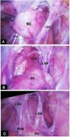
With the patient in modified right lateral decubitus, three 5 mm trocars were inserted. The cyst was identified posterior to the left subclavian artery (Fig. 2A–C); because of the intimate relation with the esophageal wall, an intra-operative upper endoscopy was performed to assist the dissection. The cystic mass was then removed en bloc, the resulting muscle defect of esophageal wall being closed with interrupted absorbable sutures; a thoracic tube was left in place through one port.

Figure 2.

(A–C): Intraoperative view.

Ao—aortic arch; BC—bronchogenic cyst; Eso—esophagus; LSA—left subclavian artery; LSV—left subclavian vein; PhN—phrenic nerve.

(0.41MB).

The postoperative course was uneventful. After removal of the thoracic tube, the child was discharged home on postoperative day 1. Histological examination revealed a uniloculated cyst lined by respiratory type epithelium with underlying fascicles of smooth muscle, respiratory-type mucous glands and cartilage, consistent with a bronchogenic cyst.

At 2-year follow-up, the child is doing well with no digestive or respiratory symptoms; the scars are almost imperceptible.

Comment

Foregut cystic malformations are rare congenital entities. Bronchogenic cysts (BC) in the mediastinum form early in fetal development from abnormal buds of tracheobronchial tree. Most of them are asymptomatic in the early stages, thus they are rare in infancy and often recognized in young adults. In fact, most diagnoses occur when the BC become infected or grow large enough to compress adjacent organs.1,2

The treatment of asymptomatic BC is not consensual. In adults, conservative management under close long-term follow-up is an option.3 In children, both symptomatic and asymptomatic cysts should be surgically excised because of risk of enlargement/compression (due to secreting mucosa or infection), erosion/perforation, bleeding, and malignant degeneration. As these lesions do not regress, it is probably more appropriate to resect asymptomatic BC not only in children but also in the young and in the healthy adult in order to prevent life threatening complications such as the present case illustrates.2,3

Drainage of symptomatic BC is a minimally invasive procedure when performed by percutaneous access under ultrasound guidance or by endobronchial ultrasound-guided fine needle aspiration (EBUS-FNA).1,4–6 The latter has been used mainly for diagnostic purpose, but several experts have applied it successfully to BC drainage1,6; unfortunately, there is no bronchoscopy with EBUS-FNA available for young children. Drainage can be used in patients who are nonsurgical candidates, but it is a less than optimal definitive treatment because the risk of infection or cyst recurrence remains. In addition to the rapid resolution of respiratory distress, the drainage should be viewed as a first step to facilitate the subsequent cyst excision due to the reduction of size and inflammation.5

Surgical excision of mediastinal BC is demanding because of vicinity and possible adhesions to surrounding vital organs, with potentially life threatening complications.5,7 In addition, some patients do not tolerate thoracoscopy well, thus thoracotomy is the procedure usually performed.5 Video-assisted thoracoscopic surgery (VATS) has been increasingly used to excise BC and is the preferred “minimally invasive technique” for most thoracic surgeons.1,5 However, a purely thoracoscopic procedure (not VATS) is even less invasive, with a more rapid recovery and better cosmesis. As the present case illustrates, it is viable despite the tight space available, particularly for suturing, a more pronounced issue in young children. So, if the patient can tolerate a “purely” thoracoscopic procedure, it should be endeavored.

In summary, this unique report demonstrates the feasibility of strict minimally invasive techniques to decompress and to excise a BC, which were crucial for the excellent outcome.

Conflicts of interest

The authors have no conflicts of interest to declare.

References
[1]
D.M. Di Bardino, K. Brenner.
Bronchogenic cyst abscess.
J Bronchol Intervent Pulmonol, 24 (2017), pp. e31-e32
[2]
B.P. Modi, C.B. Weldon.
Minimally invasive techniques for esophageal repair- foregut duplication cysts.
Adult chest surgery, 2nd ed., pp. 423-430
[3]
L. Fievet, X.B. D’Journo, J.M. Guyes, P.A. Thomas, P. De Lagausie.
Bronchogenic cyst: best time for surgery?.
Ann Thorac Surg, 94 (2012), pp. 1695-1700
[4]
S. Fernandez-Bussy, H. Inaty, I. Caviedes, G. Labarca, M.R. Vial, A. Majid.
Unusual diagnoses made by convex-probe endobronchial ultrasound-guided transbronchial needle aspiration.
Pulmonology, 24 (2018), pp. 300-306
[5]
Kawaguchi Yo, J. Hanaoka, S. Asakura, T. Fujita.
Infected bronchogenic cyst treated with drainage followed by resection.
Ann Thorac Surg, 98 (2014), pp. 332-334
[6]
V.N. Maturu, S. Dhooria, R. Agarwal.
Efficacy and safety of transbronchial needle aspiration in diagnosis and treatment of mediastinal bronchogenic cysts: systematic review of case reports.
J Bronchol Intervent Pulmunol, 22 (2015), pp. 195-203
[7]
E. Peña, M. Blanco, T. Otero.
Left main bronchus rupture during video-assisted thoracoscopic surgery resection of a bronchogenic cyst.
Rev Port Pneumol, 19 (2013), pp. 284-286
Copyright © 2021. Sociedade Portuguesa de Pneumologia
Download PDF
Pulmonology
Article options
Tools

Are you a health professional able to prescribe or dispense drugs?