

Idiopathic scoliosis is a 3-dimensional deformity of the spine, with direct effects on the thoracic cage, characterized by the lateral displacement (greater than 10°) and rotation of vertebral bodies during periods of rapid somatic growth.1 Adolescent idiopathic scoliosis (AIS) is found between the age of 10 and skeletal maturity2 and its prevalence is estimated at 2–4% in children between 10 and 16 years of age.2,3 This condition encompasses several complications including back pain, poor body image, and impaired pulmonary function.3 In fact, previous studies have shown a decreased pulmonary function in adolescents with idiopathic scoliosis,4 and an inverse correlation between scoliosis Cobb angles and pulmonary function.4 Adolescents with severe scoliosis, with Cobb angles above 45–50°, are routinely managed with spinal fusion surgery.3 In addition to the mechanical restriction to ventilation, changes in spine and thoracic cage position may alter the length of respiratory muscles influencing the ability to generate tension. Therefore, the aim of this study was to evaluate pulmonary function and respiratory muscle strength in subjects with AIS.
From November 2012 to May 2013, 12 females with AIS (15.1±1.6 years of age) and 12 age-matched controls (15.2±1.4 years of age) were enrolled in this study. The AIS group was recruited in the Paediatrics Department of Centro Hospitalar Porto-Hospital Santo António, Portugal, whereas the control group was recruited in the Porto metropolitan area. Eligible participants were those idiopathic scoliosis preoperative patients aged 10 or over, with thoracic Cobb angles of ≥40°. Exclusion criteria for this study included bronchial asthma and other pulmonary, cardiovascular or skeletal muscle problems, and previous spinal surgery. The study procedures were in accordance with the ethical standards on human experimentation. Written informed consent was obtained from parents/guardians. The Ethics Committee of the Centro Hospitalar Porto-Hospital Santo António approved the study. Lung function and respiratory muscle strength were measured before surgery. Forced expiratory volume in one second (FEV1), forced vital capacity (FVC), peak expiratory flow (PEF) and the fraction of FVC expired in one second (FEV1, FVC%) were assessed using a computerized spirometer (Spirolab III, MIR Medical International Research, Roma, Italy), according to standard methods.5 Maximal inspiratory pressure (MIP) and maximal expiratory pressure (MEP) muscle strength was measured with a digital mouth pressure meter (Micro Respiratory Muscle Analyze, CareFusion, Basingstoke, UK).6 Data were analyzed using SPSS 17.0. The normality of data distribution was tested with the Shapiro–Wilk test; the data were not normally distributed. Groups were compared by Mann–Whitney U tests. Associations between pulmonary function and respiratory muscle strength were tested with Spearman's rho test. The level of significance was set as P≤0.05.
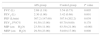
In terms of the results, the Cobb angle ranged from 42° to 62°. The AIS group presented significantly lower FEV1, FVC and PEF than the age-matched control group (Table 1). With respect to the respiratory muscle strength, both MIP and MEP were significantly higher in the control group; indeed, the median values of MIP and MEP in the control group were two times higher than those in the control group (Table 1). In the AIS group, no correlation was found between pulmonary function variables and MIP and MEP.
Spirometric and respiratory muscle strength values [median (interquartile range)].
| AIS group | Control group | P value | |
|---|---|---|---|
| FVC (L) | 2.69 (1.18) | 3.54 (0.73) | 0.005 |
| FEV1 (L) | 2.30 (1.00) | 3.42 (0.68) | 0.001 |
| PEF (L/min) | 307.2 (147.60) | 387.6 (202.2) | 0.039 |
| FEV1, FVC% | 91.50 (13.00) | 95.70 (9.00) | 0.178 |
| MIP (cmH2O) | 23.50 (11.00) | 63.50 (21.00) | <0.001 |
| MEP (cmH2O) | 29.50 (23.00) | 54.00 (17.00) | 0.008 |
FEV1, forced expiratory volume in one second; FVC, forced vital capacity; FEV1, FVC%, the fraction of FVC expired in one second; PEF, peak expiratory flow; MEP, maximal expiratory pressure; MIP, maximal inspiratory pressure.
Our results are similar to others which have shown a restrictive lung defect and impaired respiratory muscle strength7 in subjects with AIS. It is worth noting that the decrease in MIP is a major finding in the preoperative evaluation of the patient, since MIP and MEP of less than 30cm H2O augment the risk of postoperative respiratory failure.1 The inability to generate normal MIP and MEP could be a result of the chest wall deformity, which causes the respiratory muscles to work at a mechanical disadvantage.1 This study has some limitations, one of which is its small sample size. In conclusion, subjects with AIS referred to surgery showed worse pulmonary function and respiratory muscle strength than age-matched controls. From a practical standpoint, if our MIP and MEP results are corroborated by future studies, subjects with AIS could benefit from preoperative respiratory muscle strength training programmes aimed at decreasing the risk of postoperative complications.
Conflicts of interestThe authors have no conflicts of interest to declare.


