


Pulmonary hypertension (PH) is associated with poor prognosis for patients with chronic obstructive pulmonary disease (COPD). Most of the knowledge about PH in COPD has been generated at sea level, with limited information associated with high altitude (HA).
ObjectivesTo assess the prevalence and severity of PH in COPD patients living in a HA city (2,640 m).
MethodsCross-sectional study in COPD patients with forced expiratory volume in the first second / forced vital capacity ratio (FEV1/FVC) post-bronchodilator <0,7. Transthoracic echocardiography (TTE), spirometry, carbon monoxide diffusing capacity, and arterial blood gasses tests were performed. Patients were classified according to the severity of airflow limitation. PH was defined by TTE as an estimated systolic pulmonary artery pressure (sPAP) > 36 mmHg or indirect PH signs; severe PH as sPAP > 60 mmHg; and disproportionate PH as an sPAP > 60 mmHg with non-severe airflow limitation (FEV1 > 50% predicted).
ResultsWe included 176 COPD patients. The overall estimated prevalence of PH was 56.3% and the likelihood of having PH increased according to airflow-limitation severity: mild (31.6%), moderate (54.9%), severe (59.6%) and very severe (77.8%) (p = 0.038). The PH was severe in 7.3% and disproportionate in 3.4% of patients.
ConclusionsThe estimated prevalence of PH in patients with COPD at HA is high, particularly in patients with mild to moderate airflow limitation, and greater than that described for COPD patients at low altitude. These results suggest a higher risk of developing PH for COPD patients living at HA compared to COPD patients with similar airflow limitation living at low altitude.
Chronic obstructive pulmonary disease (COPD) affects around 12.6% of the world population over 40 years of age,1 and is the third most common cause of mortality worldwide and continues to rise.2 Pulmonary hypertension (PH) is a frequent complication of COPD, and its occurrence is related to increased morbidity and mortality in COPD patients.3,4 However, the increase of pulmonary artery pressure (PAP) is usually mild or moderate, and only a minor proportion of the cases develop severe PH (sPH).5-7
Although there is a clear trend indicating that the greater the severity of COPD, the greater the prevalence and severity of PH, information on the prevalence of PH in COPD patients with mild to moderate airflow limitation is scarce. Some studies using cardiac catheterization have shown a prevalence of PH in non-severe COPD patients lower than 7%.8 Using transthoracic echocardiography (TTE), prior studies have found that the prevalence of PH in patients with mild airflow limitation ranges from 0% to 25%.5,9,10
Although the development of PH in COPD patients is multifactorial, hypoxic pulmonary vasoconstriction (HPV) seems to be one central mechanism.11,12 This phenomenon is triggered by a decrease of the alveolar pressure of oxygen (PAO2) below 60 mmHg.13 So, it is reasonable to assume that chronic exposure to high altitude (HA) could be a predisposing factor for the development of PH.
It is estimated that over 140 million people in the world and 35 million in South America live at HA (> 2500 m).14,15 At HA, systolic pulmonary artery pressure (sPAP) in healthy people, as estimated by TTE, is significantly greater than that described at a low altitude.15-17 Bogotá, with around 8 million inhabitants,18 is located in the Andean Mountains at 2640 m. At this altitude, the barometric pressure (BP) is 560 mmHg, the PAO2 is around 30% lower than that at sea level, and the arterial pressure of oxygen (PaO2) in healthy adults is between 61 and 70 mmHg,19 levels close to those described as triggering HPV.13 It could be expected that patients with COPD living at this altitude may have an earlier development of PH. The objective of this study was to establish the prevalence and severity of PH in COPD patients living at HA.
Materials and methodsDesign and populationWe designed a cross-sectional analytical observational study on patients with a diagnosis of COPD and permanent residence in Bogotá. COPD was defined as forced expiratory volume in the first second / forced vital capacity ratio (FEV1/FVC) < 0.7 and exposure to a risk factor (tobacco smoke ≥ 10 packs/year and/or exposure to wood smoke > 10 years). Ambulatory patients who met the selection criteria were prospectively included. We excluded patients with exacerbations in the two months prior to recruitment; or a history of tuberculosis sequelae, bronchiectasis, silicosis, interstitial disease, chest deformity, pleural disease, or any other cardiac, respiratory, or systemic condition that could affect the sPAP, mainly primarily left ventricular failure, valvular disease, pulmonary vascular disease (primary PH or pulmonary thromboembolism), collagen disease, obesity (body mass index [BMI] > 30 Kg/m2), uncontrolled hypothyroidism, neuromuscular disease, and highly probable or known diagnosis of sleep apnea syndrome (SAS). The Sleep Apnea Clinical Score (SACS)20 and the Berlin Questionnaire21 were applied to exclude patients with a high probability of SAS. The study was approved by the Ethics Committee of the Fundación Neumológica Colombiana (approval number 200,902–14,005), and the participants signed informed consent forms.
Pulmonary function tests (PFT). Spirometry before and after bronchodilator, carbon monoxide diffusing capacity (DLCO) and arterial blood gasses (ABG) tests were performed according to the American Thoracic Society and the European Respiratory Society (ERS) standardization.22,23 Crapo's reference values were used to interpret spirometry24 and DLCO results.25 Patients were grouped by airflow-limitation severity according to the Global Initiative for Chronic Obstructive Lung Disease (GOLD).26
TTE. The exams were performed by two experienced cardiologists using a Philips Sonos 5500® with a 3.2 MHz transducer. Specific views included the parasternal long- and short-axis; apical 4, 2, and 3 chamber, and subcostal views, which allowed to evaluate the respiratory collapse of the inferior vena cava. The sPAP was estimated by measuring the tricuspid regurgitation velocity (TRV), assessed from multiple views, and searching for the best envelope and maximum velocity. The highest transvalvular velocity was used for calculating the TRV.27 Saline solution was administered through a peripheral intravenous line to magnify the signal and allow measurement in patients with trivial tricuspid regurgitation.28 Using Doppler, we also evaluated the presence of indirect PH signs, such as the right ventricular outflow doppler (RVOT) acceleration time and the late diastolic pulmonary regurgitation velocity, that have been used as PH indicators in subjects whose tricuspid regurgitation could not be easily evaluated.27,29,30 Echocardiographers were blind to the results of the PFTs and to the other operator's interpretation.
Definition ofPH and sPH. According to Chemla's evaluation of various empirical formulas for estimating mean pulmonary artery pressure in adults by using sPAP,31 and the Cologne Consensus Conference 2011 for updating the European Society of Cardiology and the ERS Guidelines on Diagnosis and Treatment of PH,32 we defined PH as a sPAP > 36 mmHg, estimated by TTE. For subjects in whom the flow of tricuspid regurgitation could not be measured, we used the aforementioned indirect signs. RVOT acceleration time <105 ms and early diastolic pulmonary regurgitation velocity > 2.2 m/s were considered markers of raised PAP.27,30 For the comparison of our results with the literature, we defined sPH as an sPAP > 60 mmHg,33 which would be equivalent to a mean PAP of 40 mmHg according to the Chemla equation,34 and disproportionate PH as a sPAP > 60 mmHg with non-severe airflow limitation (FEV1> 50% predicted).35
Statistical analysisThe normality of variables was tested using the Kolmogorov-Smirnov test. Continuous variables were expressed as mean ± standard deviation (SD) or median and interquartile ranges and qualitative variables as proportions. To assess differences between the groups with and without PH, we used Chi-square tests, unpaired t-tests, and nonparametric Mann-Whitney tests. For differences in PH prevalence between GOLD stages, we used Chi-square tests. Intra-observer agreement of sPAP measurements was blindly assessed in half of the echocardiograms using Lin's correlation coefficient of agreement. A correlation matrix was created to relate sPAP and PFT. The associations of the presence of PH with sex, age, BMI, the severity of airflow limitation, DLCO, and ABG variables were examined in a multivariate logistic regression model. The oxygen arterial pressure (PaO2) and carbon dioxide arterial pressure (PaCO2) were distributed dichotomously, using as cut-off levels the normality limits for the altitude of Bogotá.19 Variables with a value of p < 0.2 in the univariate analysis or with a possible physiological relationship with PH were included in the final model. Adjusted odds ratios (OR) with their corresponding 95% confidence intervals (CI) were calculated. Analyses were performed using the statistical software SPSS 15.0.
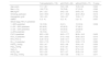
ResultsA total of 176 patients, 77% male, were included. Table 1 shows the baseline characteristics of participants.
Characteristics of COPD patients with and without pulmonary hypertension.
Data presented as mean ± SD, median (P25-P75) or N (%). P= differences between patients with and without pulmonary hypertension. PH: pulmonary hypertension; BMI: body mass index; mMRC: modified Medical Research Council scale; GOLD: Global Initiative for Chronic Obstructive Lung Disease; FEV1: forced expiratory volume in the first second; FVC: forced vital capacity; DLCO: carbon monoxide diffusing capacity; PaCO2: arterial partial pressure of carbon dioxide; PaO2: arterial partial pressure of oxygen; SaO2: arterial oxygen saturation; HCO3: bicarbonate; P(A-a)O2: alveolar-arterial oxygen gradient.
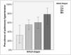
The global prevalence of PH was 56.3% and was higher in patients with more severe GOLD stages (GOLD 1: 31.6%; GOLD 2: 54.9%; GOLD 3: 59.6%; GOLD 4: 77.8%; p = 0.038) (Figure 1).

In 93.8% of patients with PH, the diagnosis was established using the sPAP > 36 mmHg criteria. Thirteen patients (7.3%) presented sPH with an estimated sPAP greater than 60 mmHg, and six of these patients (3.4% of the total) had mild or moderate airflow limitation.
Compared to patients without PH, patients with PH had lower FEV1, FEV1/FVC, PaO2, and oxygen arterial saturation (SaO2) and higher PaCO2 (Table 1). We found a weak but statistically significant inverse correlation between sPAP and FEV1%, DLCO%, SaO2, and PaO2 and a direct correlation between sPAP and P(A-a)O2 and PaCO2 (Table 2). In the multivariate analysis, PaO2 < 55 mmHg and very severe airflow limitation (FEV1 < 30% predicted) maintained significant associations with pH (Table 3).
Correlation between sPAP with pulmonary function tests and arterial blood gasses.
sPAP: systolic pulmonary artery pressure; FVC: forced vital capacity; FEV1: forced expiratory volume in the first second; DLCO: carbon monoxide diffusing capacity; PaO2: arterial partial pressure of oxygen; SaO2: arterial oxygen saturation; PaCO2: arterial partial pressure of carbon dioxide; P(A-a)O2: alveolar-arterial oxygen gradient.
R= Pearson coefficient.
Risk factors for pulmonary hypertension - Multivariate analysis.
GOLD: Global Initiative for Chronic Obstructive Pulmonary Disease; PaCO2: arterial partial pressure of carbon dioxide; PaO2: arterial partial pressure of oxygen; OR: odds ratio.
In the 89 patients in whom it was possible to measure tricuspid regurgitation, concordance between the first and second estimations of sPAP was 0.905 (p < 0.001).
DiscussionOur study shows that patients with COPD living at HA have a high prevalence of PH (56.3%). The risk factors associated with PH were the PaO2 and GOLD stage 4. The high prevalence of PH (31.6%) in patients with mild to moderate airflow limitation found in our study suggest a higher risk of developing pH for COPD patients living at HA compared to COPD patients with similar airflow limitation living at low altitude. In addition, we also found a greater frequency of sPH than that described at low altitude.
Previous studies on COPD patients with mild airflow limitation have shown a prevalence of PH from 0% to 16.7%.5,10 Although one study showed a higher prevalence (25%),9 mild airflow limitation was defined as FEV1 > 60% predicted, which included subjects with more advanced diseases. In our COPD population residing at HA (2640 m), patients with moderate or severe airflow limitation also had a greater prevalence of PH than that described at low altitude, except in one study5 that showed similar results. In subjects with very severe airflow limitation, the prevalence of PH found in our study was similar to that described at sea level.5,9,10
The distinctive factor of our study is the permanent residency of the patients at HA (2640 m), where PaO2 and PAO2, in healthy people over 40 years old, are close to 60 mmHg,19 a level at which the onset of HPV has been documented.13 At this level, small regional or general variations in alveolar ventilation and ventilation/perfusion ratio could trigger HPV and potentially generate PH. We propose that in patients with COPD, residency at HA could favor the early development of permanent or intermittent hypoxaemia at rest or during sleep or exercise,36 which by itself or in interaction with genetic factors,37,38 sleep disorders,39 or lung and systemic inflammation40-42 could promote the development of PH.
In healthy people resident above 3600 m, it has been described higher sPAP compared to residents at sea level.16,17 This finding confirms that living at a HA is a factor directly influencing the pressure in the pulmonary artery, even in people without respiratory diseases.
As previously described, the mean pressure in the pulmonary artery is higher in residents at HA compared to those at sea level, which is accompanied by changes in the right ventricle.43 Interestingly, the mean of sPAP in our population (43.8 mmHg ± 15.7 mmHg) was very similar to that previously described in COPD patients residing at 1768 m (43.1 mmHg ± 11.8 mmHg), which further indicates the influence of altitude on the pulmonary vasculature.44 In this sense, a high prevalence of PH in patients with idiopathic pulmonary fibrosis at HA has been recorded.45
We also found a high prevalence of sPH (7.3%) in our patients. This prevalence is greater than that described in patients with severe airflow limitation using cardiac catheterization.7,33,46,47 Studies using TTE have found similar6,48 or lower5 prevalence of sPH, although one of these studies was performed on patients with recent exacerbation,48 a known condition associated with increased PAP.49,50 Interestingly, we found that 3.4% of patients had disproportionate PH (sPH with non-severe airflow limitation). Most cases of patients with COPD and sPH may have additional conditions responsible for increased PAP and pH.31 In our study we reasonably excluded patients with those conditions. Although most patients with pH had non-severe increases in sPAP, it is not clear if at HA, this level of PH constitutes a risk factor for mortality, as previously described in the general population, even with borderline sPAP values.51,52
Similar to what has been previously documented,9,48,53 we found significant correlations between PH and some physiological variables such as FEV1, DLCO, PaCO2, PaO2, and SpO2. However, these variables did not completely explain the changes of sPAP, suggesting that other variables, not evaluated herein, could play an important role in increased PAP in COPD patients. Some conditions previously related to increased PAP, such as weight49 and heart function,54 were reasonably controlled by excluding patients with BMI ≥ 30 Kg/m2 or significant heart dysfunction. We did not evaluate biomarkers of systemic inflammation such as C-reactive protein (CRP) and tumoral necrosis factor alpha (TNF-α) that have previously been associated with PH in patients with COPD.55
SAS leads to a longer desaturation time in COPD patients at HA when compared with those living at sea level.39,56 We excluded patients with a previous diagnosis or high suspicion of SAS.
Our study suggests a notable role of HA and its consequent lower PAO2 and PaO2 on the frequent development of pH in patients with COPD. In these cases, PH can be a treatable trait, since in COPD patients with hypoxaemia57 or with pulmonary arterial or distal chronic thromboembolic pulmonary hypertension presenting with mild resting hypoxaemia and exercise-induced oxygen desaturation,58 oxygen therapy can acutely improve exercise tolerance57,58 and long-term survival.59,60 Patients with sPH and disproportionate PH constitute a particular group that should be evaluated by experts in PH.11,54,61
To the best of our knowledge, this is the largest study evaluating the prevalence of PH in stable COPD patients living at HA. A strength of the study is the prospective inclusion of subjects with confirmed COPD and all degrees of airflow limitation, among whom we reasonably discarded any with coexisting pathologic conditions that could contribute to PH. TTE were performed in a single center by a small number of highly specialized cardiologists, using rigorous criteria, and with a high concordance between observers.
Our study has some limitations. Because our institution is a reference center, our sample has a lower proportion of patients with mild airflow limitation than that described in other studies.62 Hence, the high prevalence of PH found in our study could not be reliably extrapolated to the general population with COPD living at similar altitudes. We also do not have information on COPD exacerbations in the year prior to study entry to classify our patients according to GOLD classification, beyond the degree of airflow limitation, which may constitute a weakness since a higher prevalence of PH has been previously reported in groups C and D.6
Another limitation is the use of TTE for evaluating PH. Although this exam is not the gold standard for confirming PH, it is a widely used tool in clinical practice given its low cost, availability, and safety.63,64 In addition,PH estimated by TTE has been related to clinically important outcomes such as exercise tolerance in COPD.65,66
A major limitation of TTE for the evaluation of PAP in COPD patients is that it is frequently not possible to detect tricuspid regurgitation to estimate sPAP.9,67,68 However, in our study, we found a high rate of assessable tricuspid regurgitation flow (89%). Some studies have found that sPAP estimated by TTE has a significantly high level of agreement with catheterization measurements.67,69 We used an estimated sPAP > 36 mmHg to define pH, a cut-off point that has been described as having high sensitivity, specificity, and good negative predictive value for the diagnosis of PH.31,32,67 We used indirect signs and contrast with shaken saline for improving the operating characteristics of the TTE, as has been described.27,28,63
In conclusion, the prevalence of PH in patients with COPD living in Bogotá, a city located at HA (2640 m), is high and greater than that described at sea level, particularly in patients with less severe airflow limitation, suggesting a higher risk of developing PH for COPD patients living in HA compared to COPD patients with similar airflow limitation living at low altitudes. Likewise, the prevalence of sPH and disproportionate PH was also higher at HA.
Cra. 13b #161–85, Bogotá, Colombia
Cra. 13b #161–85, Bogotá, Colombia.
Cll. 163a #13B-60, Bogotá, Colombia.


- Home
- All contents
- About the journal
- Metrics
- Open access