


Insufficient cough strength has a major role in extubation and decannulation outcomes.
Cough capacity can be easily evaluated by measuring flows during coughing. Values vary depending on whether cough flows are measured through the mouth or through a tracheostomy or endotracheal tube. It is important to standardize these measurements and start using them routinely in the extubation and decannulation processes. Values of cough peak flow >160 L/min measured at the mouth or a value of cough PEF >60 L/min measured at the endotracheal tube suggest successful decannulation or extubation.
Insufficient cough strength plays a major role in failed extubation/decannulation in patients with high level spinal cord injury, primary neuromuscular disorders or ICU-acquired weakness.1, 2
Evidence-based assessments in the ventilator discontinuation process3 suggest evaluation of cough strength in patients passing a spontaneous breathing trial, however this procedure has not yet moved into clinical practice.
Extubation failure rate ranges from 10% to 20% of extubations and translates into higher mortality compared with successful extubation.4
Decannulation failure ranges from 2%,5 in acute brain injury patients in rehabilitation facilities, to 32.4%6 in patients with neuromuscular ventilator insufficiency (predominantly spinal cord injuries) admitted to a ventilator unit. More recently in a prospective study, Choate et al.7 report decannulation failure rate of 4.8%. Comparison between these studies is difficult because of differences in definitions of decannulation failure, lack of consistency in weaning and decannulation protocols, and differing patient characteristics.
Patients who are decannulated have better survival rates. O’Connor et al.8 examined the process of decannulation in patients transferred to a long-term acute care hospital. Decannulation was successful in 35% of patients at a median of 45 days following tracheostomy. Patients who failed decannulation had a tracheostomy tube placed earlier and had a shorter stay in the referring acute care hospital. At 3.5 years, 35% of all patients and 62% of decannulated patients were alive.
Where decannulation fails the majority require simple recannulation but others (37.5%) may require translaryngeal intubation,7 and readmission to the ICU.
Cough capacity can be easily evaluated by measuring flows during coughing.1, 6 If cough flows are measured through the mouth (with an active glottis) we can call it a true cough peak flow (CPF)6 whereas if measured through a tracheostomy or an endotracheal tube it is more a peak expiratory flow (PEF), because of bypassing the glottis.1
Cough PEF values during extubation (see also Table 1 )In a study by Smina et al.1 cough PEF was the strongest predictor of extubation outcome for patients without neuromucular disorders. The mean PEFs were significantly lower in unsuccessful extubations compared with successful extubations (64.2 ± 6.8 L/min vs 81.9 ± 2.7 L/min, p = 0.03) (Table 1).
Table 1. Summary of studies.
| Article | Number of patients | Diagnosis | Measurement via mouth/trach/ET | Device | Outcome |
| 1 | 95 | Critically ill | ET | Aztech | Extubation failure, mortality |
| 2 | 150 | Critically ill | ET | Respiratory mechanics monitor | Extubation failure |
| 6 | 49 | Neuromuscular | Mouth | HealthScan | Decannulation failure |
| 9 | 130 | Critically ill | ET | Piko-1 | Extubation failure |
| 10 | 32 | Neurosurgical | Trach | Piko-1 | Decannulation failure |
| 11 | 88 | Critically ill | ET | Aztech | Extubation failure |
| 12 | 125 | Burn | ET | Mini-Wright | Extubation failure |
| 13 | 61 | Neuromuscular | Mouth | Access | Post-decannulation, vital capacity |
| 14 | 26 | Neuromuscular | Mouth | Mini-Wright | Pre- vs post-decannulation value, decannulation failure |
| 15 | 151 | Prolonged Weaning or Neurological | Trach | Mini-Wright | Time to decannulation |
In the Pascal Beuret et al. study,9 which included ICU patients who had successfully passed the spontaneous breathing trial, the optimal cough PEF value to predict extubation failure was >35 L/min.
In patients unable to cooperate voluntarily cough PEF cannot be evaluated. Involuntary cough may be assessed either by instilling normal saline2 or by advancing a suctioning catheter through the patient's tracheostomy tube to induce a cough.10 Although not compared directly two different studies using voluntary11 and involuntary cough PEF2 have come out with approximately the same cut-off point (60 versus 58.5 L/min respectively)
In the Salam et al. study11 Cough PEFs were measured in all but five patients who were unable to understand instructions or could not attempt a cough. For the remaining 83 patients who attempted to cough, the Cough PEFs ranged from 10 to 200 L/min; 41% of the measurements were 60 L/min or less.
The study of Smailes12 demonstrated that patients with a cough PEF of ≤60 L/min are nine times as likely to fail extubation as those with a CPF > 60 L/min. Smina et al. and Salam et al.1, 11 also identified a cough PEF threshold of 60 L/min. The study by Beuret et al.9 identified a much lower Cough PEF threshold, which might be due to different patient populations or characteristics of the device used to measure cough.
In effect the Cough PEF measured in intubated patients may be considered a “huff” or, perhaps, a peak expiratory flow maneuver and not a true peak cough flow.
Cough peak flow (CPF) and cough PEF values during decannulation (see also Table 1 )Bach and Saporito were the first to use CPF to predict decannulation failure.6 They proposed providing maximal insufflations together with an abdominal thrust timed to glottic opening (manually assisted coughing). In this way assisted CPF of at least 160 L/min (immediately after extubation or decannulation) predicted successful extubation or decannulation.
More recently the same authors13 challenged the cut-off stating that with great experience in noninvasive management it was possible to extubate and decannulate patients even with CPF < 160 L/min; however, they warned that the less the upper airway patency as suggested by low CPF or mechanical in-exsufflation (MIE) flows, the less effective is MIE in keeping the airways clear after extubation or decannulation and the greater the risk of failing noninvasive management.
McKim et al.14 measured CPF through the mouth in tracheostomized patients with the cuffs deflated, and cannulas capped. In this study, spontaneous CPF through the mouth in tracheostomized neuromuscular patients was about 35 L/min lower than post-decannulation spontaneous CPF. Those authors proposed that with the gain achieved post-decannulation, a spontaneous CPF value of 130 L/min may still anticipate a successful decannulation.
Hernandez et al.15 in 151 medical-surgical ICU patients measured cough PEF through the tracheostomy tube and showed that values were higher with the cuff deflated. Moreover they demonstrated that low PEF (percentile 33) was negatively associated with decannulation. Mean PEF in the 87 patients with prolonged weaning was 118 ± 77 L/min.
Cough flow values: through the tube or through the mouth?Peak flows through an endotracheal tube are bound to be lower than those in decannulated patients because intubated patients cannot close their glottis, thereby limiting the pressure generated when attempting to cough. Thus, the reason for the lower threshold values described in the studies measuring CPF in intubated patients is a result of the different procedures as these patients perform a “glotic-free cough PEF.”
Set-up to measure cough PEF in patients with an endotracheal tubeThe peak flow meter/pneumotachograph should be connected to the proximal tip of the tracheal tube via a bacterial/viral filter. The patient, who should be positioned in the semi-recumbent position, should then be instructed to inspire deeply through a three-way connector positioned between the proximal tip of the tracheal tube and the peak flow meter. The external port of the connector should then be occluded, and the patient instructed to cough as strongly as possible through the tracheal tube.
Alternatively, involuntary Cough PEF can be induced by removing the T-piece and dripping 2 mL of normal saline into the endotracheal tube at the end-inspiratory point, with the patient in a head-up position at 45° from the horizontal. The end-inspiratory point is chosen to ensure that a full inspiration has occurred.
The T-piece and flow sensor should be reconnected to the endotracheal tube as soon as possible. The patients should be continuously observed until their breathing becomes smooth and regular. The maximum expiratory flow value detected should be recorded as the involuntary cough PEF.2
Set-up to measure cough flows in patients with a tracheostomy tubeMeasurement through the cannula ( Fig. 1 )In uncooperative patients (e.g. neurosurgical patients), the patient should be in the supine position with the head of the bed elevated at 30°. Patients with a cuffed tracheotomy tube should have the cuff deflated before measurements are made. Suctioning above the cuff should be done before the cuff is deflated. Oxygen saturation and signs of respiratory distress should be monitored during the measurement process. The cough PEF measurement procedure should be stopped immediately if any of the following occur: respiratory rate greater than 35/min; oxygen saturation, as measured by pulse oximetry, less than 90%; heart rate above 140/min or an increase of more than 20% above resting levels. With sterile conditions, a standardized proportion (12 cm) of a 10F suction catheter should be introduced through the suction port of the swivel elbow connector. The swivel connector with the suction catheter partially inserted is then attached to the patient's tracheotomy tube, which is in turn connected to a viral/bacterial respiratory filter allowing a pneumotachograph-calibrated electronic peak flow meter (Figure 1).
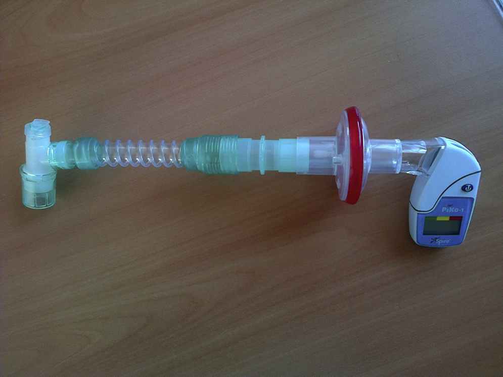
Figure 1. Set-up of an electronic peak flow meter (PiKo I) for measuring cough PEF through the tracheal cannula.
Measurement through the mouth ( Fig. 2 )For cooperative patients the CPF measurements can be done with cuffs deflated, tracheostomy cannulas capped and measured through the mouth14 (Figure 2).
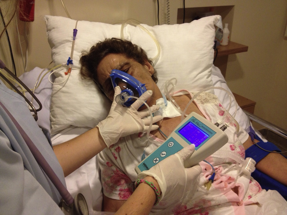
Figure 2. Set-up of a hand held respiratory mechanics monitor for measuring PCF through the mouth.
Patients should be asked to cough via an oronasal interface into a peak flow meter with the tracheostomy tube covered and cuff deflated.13 If a spontaneous CPF of 160 L/min is not achieved then an assisted CPF should be determined by removing the tube, covering the ostomy while ventilating the lungs by mouth piece non-invasive ventilation as needed, and getting the patient to air stack consecutive ventilator-delivered volumes via a mouth piece to a deep lung volume and then coughing into a peak flow meter as an abdominal thrust is applied (called manually assisted CPF, that is the value following lung volume recruitment and manual abdominal trust).
Alternatively, to measure CPF after lung volume recruitment, breaths can be stacked to approximate the maximum insufflation capacity.14 To achieve a maximum insufflation capacity, the patient is instructed to inhale fully, hold his/her breath, and then place the lips tightly around a mouthpiece through which consecutive volumes of air are delivered using a manual resuscitation bag and held by a closed glottis. The patient is then asked to cough and the flows are measured (called lung volume recruitment CPF).
Measurements of cough flows: with mechanical peak flow meter, electronic peak flow meter or pneumotachograph? (see also Table 1 )There is a great variability of devices used in the studies: from a classical mini-Wright Peak flow meter,12, 14, 15 an Aztech Peak flow meter,1, 11 an Access Peak flow meter,6, 13 to an electronic peak flow meter9, 10 or a respiratory mechanics monitor.2
Sancho et al.16 compared CPF measurements with a mechanical peak flow meter (asmaPLAN, Vitalograph, Ennis, Ireland) and pneumotachograph in healthy volunteers and spontaneously breathing neuromuscular disorders patients. In their study PCF measurements made with the peak flow meter were reproducible and reasonably accurate when flows were >270 L/min. However, the authors have warned that caution is needed in clinical practice at lower PCF because of the tendency of this portable device to overestimate the lower flows.16
Electronic PEF meters (like the PiKo-1) can measure PEF in the range of 15 to 999 L/min with a 1 L/min resolution, an accuracy of 6.5%, and a better agreement with pneumotachograph compared to mechanical PEF meters. Due to its low cost and storage capacity it could be a good alternative to the more expensive pneomotachograph.17
Analysis of cough flow–volume curvesPresence of transients of peak flow during cough flow-volume maneuvers may suggest also cough efficiency.18 In fact in a prospective study including 53 spontaneously breathing patients with Amyotrophic lateral sclerosis, the absence of cough spikes in the flow-volume loops was related to an increased mortality.19 So analysis of cough flow–volume curves with the use of a pneumotachograph may add to the analysis of the absolute CPF values.
ConclusionsCough flows should become the preferred method of assessing cough strength in patients for whom extubation or decannulation is being planned. Only when the measurement is obtained with an active glottis should we call it cough peak flow, otherwise it should be termed cough PEF.
Although cut-off values for high-risk patients have been defined, different methodologies (set-up and measuring devices) and patient populations involved in the studies suggest some caution in interpreting absolute values. However as a rule of thumb a value of cough peak flow >160 L/min measured at the mouth or a value of endotracheal cough PEF >60 L/min measured suggests that they are good candidates for decannulation or extubation.
Due to lower cough flow values in this setting, use of more accurate devices like electronic PEF meters should be recommended.
Conflict of interestThe authors have no conflicts of interest to declare.
Received 14 December 2014
Accepted 16 December 2014
Corresponding author. jcwinck@mail.telepac.pt


