array:20 [
  "pii" => "X0873215915365019"
  "issn" => "08732159"
  "doi" => "10.1016/j.rppnen.2015.06.003"
  "estado" => "S300"
  "fechaPublicacion" => "2015-09-01"
  "documento" => "article"
  "licencia" => "http://www.elsevier.com/open-access/userlicense/1.0/"
  "subdocumento" => "fla"
  "cita" => "Rev Port Pneumol. 2015;21:284-5"
  "abierto" => array:3 [
    "ES" => true
    "ES2" => true
    "LATM" => true
  ]
  "gratuito" => true
  "lecturas" => array:2 [
    "total" => 4933
    "formatos" => array:3 [
      "EPUB" => 244
      "HTML" => 3766
      "PDF" => 923
    ]
  ]
  "itemSiguiente" => array:16 [
    "pii" => "X0873215915365027"
    "issn" => "08732159"
    "doi" => "10.1016/j.rppnen.2015.05.002"
    "estado" => "S300"
    "fechaPublicacion" => "2015-09-01"
    "documento" => "article"
    "licencia" => "http://www.elsevier.com/open-access/userlicense/1.0/"
    "subdocumento" => "fla"
    "cita" => "Rev Port Pneumol. 2015;21:285-6"
    "abierto" => array:3 [
      "ES" => true
      "ES2" => true
      "LATM" => true
    ]
    "gratuito" => true
    "lecturas" => array:2 [
      "total" => 3761
      "formatos" => array:3 [
        "EPUB" => 244
        "HTML" => 2578
        "PDF" => 939
      ]
    ]
    "en" => array:8 [
      "idiomaDefecto" => true
      "titulo" => "Misconceptions in the assessment of cough peak flow measurements for extubation or decanulation protocols"
      "tienePdf" => "en"
      "tieneTextoCompleto" => "en"
      "paginas" => array:1 [
        0 => array:2 [
          "paginaInicial" => "285"
          "paginaFinal" => "286"
        ]
      ]
      "contieneTextoCompleto" => array:1 [
        "en" => true
      ]
      "contienePdf" => array:1 [
        "en" => true
      ]
      "autores" => array:1 [
        0 => array:2 [
          "autoresLista" => "M. Chiou, J.R. Bach, M.R. Gonçalves, L. Vudayagiri"
          "autores" => array:4 [
            0 => array:2 [
              "Iniciales" => "M."
              "apellidos" => "Chiou"
            ]
            1 => array:2 [
              "Iniciales" => "J.R."
              "apellidos" => "Bach"
            ]
            2 => array:2 [
              "Iniciales" => "M.R."
              "apellidos" => "Gonçalves"
            ]
            3 => array:2 [
              "Iniciales" => "L."
              "apellidos" => "Vudayagiri"
            ]
          ]
        ]
      ]
    ]
    "idiomaDefecto" => "en"
    "EPUB" => "https://multimedia.elsevier.es/PublicationsMultimediaV1/item/epub/X0873215915365027?idApp=UINPBA00004E"
    "url" => "/08732159/0000002100000005/v0_201604141143/X0873215915365027/v0_201604141143/en/main.assets"
  ]
  "itemAnterior" => array:16 [
    "pii" => "X0873215915365000"
    "issn" => "08732159"
    "doi" => "10.1016/j.rppnen.2015.05.003"
    "estado" => "S300"
    "fechaPublicacion" => "2015-09-01"
    "documento" => "article"
    "licencia" => "http://www.elsevier.com/open-access/userlicense/1.0/"
    "subdocumento" => "fla"
    "cita" => "Rev Port Pneumol. 2015;21:282-4"
    "abierto" => array:3 [
      "ES" => true
      "ES2" => true
      "LATM" => true
    ]
    "gratuito" => true
    "lecturas" => array:2 [
      "total" => 3562
      "formatos" => array:3 [
        "EPUB" => 238
        "HTML" => 2449
        "PDF" => 875
      ]
    ]
    "en" => array:9 [
      "idiomaDefecto" => true
      "titulo" => "Late onset congenital cystic adenomatous malformation associated with intra-lobar pulmonary sequestration"
      "tienePdf" => "en"
      "tieneTextoCompleto" => "en"
      "paginas" => array:1 [
        0 => array:2 [
          "paginaInicial" => "282"
          "paginaFinal" => "284"
        ]
      ]
      "contieneTextoCompleto" => array:1 [
        "en" => true
      ]
      "contienePdf" => array:1 [
        "en" => true
      ]
      "resumenGrafico" => array:2 [
        "original" => 0
        "multimedia" => array:8 [
          "identificador" => "fig1"
          "etiqueta" => "Fig. 1"
          "tipo" => "MULTIMEDIAFIGURA"
          "mostrarFloat" => true
          "mostrarDisplay" => false
          "copyright" => "Elsevier España"
          "figura" => array:1 [
            0 => array:4 [
              "imagen" => "320v21n05-90436500fig1.jpg"
              "Alto" => 2363
              "Ancho" => 1795
              "Tamanyo" => 2143579
            ]
          ]
          "descripcion" => array:1 [
            "en" => "(A) The first chest CT performed, showing a heterogeneous posteromedial consolidation in the left lower lobe comprising multiple cysts. (B) The second chest CT showing posteromedial hepatization of the left lower field, compatible with superimposed infection. (C) CT angiography showing the anomalous artery arising from the thoracic aorta. (D) CT angiography images showing the venous drainage (arrow) to the pulmonary veins. (E¿F) 3D CT reconstructions showing the anomalous artery."
          ]
        ]
      ]
      "autores" => array:1 [
        0 => array:2 [
          "autoresLista" => "E. Silva, J. Vale, A. Reis"
          "autores" => array:3 [
            0 => array:2 [
              "Iniciales" => "E."
              "apellidos" => "Silva"
            ]
            1 => array:2 [
              "Iniciales" => "J."
              "apellidos" => "Vale"
            ]
            2 => array:2 [
              "Iniciales" => "A."
              "apellidos" => "Reis"
            ]
          ]
        ]
      ]
    ]
    "idiomaDefecto" => "en"
    "EPUB" => "https://multimedia.elsevier.es/PublicationsMultimediaV1/item/epub/X0873215915365000?idApp=UINPBA00004E"
    "url" => "/08732159/0000002100000005/v0_201604141143/X0873215915365000/v0_201604141143/en/main.assets"
  ]
  "en" => array:10 [
    "idiomaDefecto" => true
    "titulo" => "Yellow bronchoscopy: A severe case of tracheal and bronchial Jaundice"
    "tieneTextoCompleto" => true
    "paginas" => array:1 [
      0 => array:2 [
        "paginaInicial" => "284"
        "paginaFinal" => "285"
      ]
    ]
    "autores" => array:1 [
      0 => array:4 [
        "autoresLista" => "A. Gonçalves, C. Ribeiro, A. Oliveira"
        "autores" => array:3 [
          0 => array:4 [
            "Iniciales" => "A."
            "apellidos" => "Gonçalves"
            "email" => array:1 [
              0 => "ana_f_goncalves@hotmail.com"
            ]
            "referencia" => array:2 [
              0 => array:2 [
                "etiqueta" => "<span class="elsevierStyleSup">a</span>"
                "identificador" => "affa"
              ]
              1 => array:2 [
                "etiqueta" => "<span class="elsevierStyleSup">&#42;</span>"
                "identificador" => "cor1"
              ]
            ]
          ]
          1 => array:3 [
            "Iniciales" => "C."
            "apellidos" => "Ribeiro"
            "referencia" => array:1 [
              0 => array:2 [
                "etiqueta" => "<span class="elsevierStyleSup">a</span>"
                "identificador" => "affa"
              ]
            ]
          ]
          2 => array:3 [
            "Iniciales" => "A."
            "apellidos" => "Oliveira"
            "referencia" => array:1 [
              0 => array:2 [
                "etiqueta" => "<span class="elsevierStyleSup">a</span>"
                "identificador" => "affa"
              ]
            ]
          ]
        ]
        "afiliaciones" => array:1 [
          0 => array:3 [
            "entidad" => "Pulmonology Department, Centro Hospitalar Vila Nova de Gaia/Espinho, Portugal"
            "etiqueta" => "<span class="elsevierStyleSup">a</span>"
            "identificador" => "affa"
          ]
        ]
        "correspondencia" => array:1 [
          0 => array:3 [
            "identificador" => "cor1"
            "etiqueta" => "<span class="elsevierStyleSup">&#42;</span>"
            "correspondencia" => "Corresponding author. ana_f_goncalves@hotmail.com"
          ]
        ]
      ]
    ]
    "resumenGrafico" => array:2 [
      "original" => 0
      "multimedia" => array:8 [
        "identificador" => "fig1"
        "etiqueta" => "Fig. 1"
        "tipo" => "MULTIMEDIAFIGURA"
        "mostrarFloat" => true
        "mostrarDisplay" => false
        "copyright" => "Elsevier Espa&#241;a"
        "figura" => array:1 [
          0 => array:4 [
            "imagen" => "320v21n05-90436501fig1.jpg"
            "Alto" => 1504
            "Ancho" => 1500
            "Tamanyo" => 670326
          ]
        ]
        "descripcion" => array:1 [
          "en" => "&#40;A&#41; Epiglottis&#59; &#40;B&#41; Vocal cords&#59; &#40;C&#41; Carina&#59; &#40;D&#41; Truncus intermedius&#46;"
        ]
      ]
    ]
    "textoCompleto" => "<p class="elsevierStylePara">Dear Editor&#44;</p><p class="elsevierStylePara">Jaundice&#44; which can be caused by a very wide variety of conditions&#44; results from the increased levels of bilirubin in the blood&#46; Due to the affinity of bilirubin to elastin&#44; jaundice is best detected by examining the skin and sclera&#44; but can also be found in others elastin-rich tissues such as skin&#44; sclera&#44; intima of the vessel wall and ligaments&#46;</p><p class="elsevierStylePara">We present the case of a 68-year-old nonsmoker male who was referred to the emergency department for evaluation of 1-week history of jaundice&#44; light-colored stools&#44; dark urine&#44; anorexia and weight loss&#46; Blood work showed an elevated direct bilirubin 9&#46;34&#160;mg&#47;dL &#40;normal&#44; 0&#8211;1&#160;mg&#47;dL&#41; and he was diagnosed with a carcinoma of the pancreas head&#46;</p><p class="elsevierStylePara">Bronchoscopy was performed during a pulmonary nodule investigation and revealed a nodule on the left side of the epiglottis and severe vocal cords and bronchial tree jaundice &#40;<a href="&#35;f0005" class="elsevierStyleCrossRefs">Fig&#46; 1</a>&#41;&#46;</p><a name="f0005" class="elsevierStyleCrossRefs"></a><p class="elsevierStylePara"><img src="320v21n05-90436501fig1.jpg" alt="&#40;A&#41; Epiglottis&#59; &#40;B&#41; Vocal cords&#59; &#40;C&#41; Carina&#59; &#40;D&#41; Truncus intermedius&#46;"></img></p><p class="elsevierStylePara">Fig&#46; 1&#46; &#40;A&#41; Epiglottis&#59; &#40;B&#41; Vocal cords&#59; &#40;C&#41; Carina&#59; &#40;D&#41; Truncus intermedius&#46;</p><p class="elsevierStylePara">Bronchoscopy is not a frequently performed exam in patients with such high levels of bilirubin in the blood&#46; In the present case bronchoscopy was performed due to the nodule location&#44; making it difficult to access through transthoracic lung biopsy and due to the need for pulmonary metastasis exclusion in order to get a correct staging&#46;</p><p class="elsevierStylePara">The high amount of elastin present in the airways makes it possible to observe a fine yellow coloration characteristic of jaundice in the bronchial tree&#44; as observed in these endoscopic images of rare beauty and iconographic interest&#46;</p><a name="sec0005" class="elsevierStyleCrossRefs"></a><span class="elsevierStyleSectionTitle">Conflict of interest</span><p class="elsevierStylePara">The authors have no conflicts of interest to declare&#46;</p><p class="elsevierStylePara">Corresponding author&#46; ana&#95;f&#95;goncalves&#64;hotmail&#46;com</p>"
    "pdfFichero" => "320v21n05a90436501pdf001.pdf"
    "tienePdf" => true
    "multimedia" => array:2 [
      0 => array:8 [
        "identificador" => "fig1"
        "etiqueta" => "Fig. 1"
        "tipo" => "MULTIMEDIAFIGURA"
        "mostrarFloat" => true
        "mostrarDisplay" => false
        "copyright" => "Elsevier Espa&#241;a"
        "figura" => array:1 [
          0 => array:4 [
            "imagen" => "320v21n05-90436501fig1.jpg"
            "Alto" => 1504
            "Ancho" => 1500
            "Tamanyo" => 670326
          ]
        ]
        "descripcion" => array:1 [
          "en" => "&#40;A&#41; Epiglottis&#59; &#40;B&#41; Vocal cords&#59; &#40;C&#41; Carina&#59; &#40;D&#41; Truncus intermedius&#46;"
        ]
      ]
      1 => array:6 [
        "identificador" => "fig2"
        "tipo" => "MULTIMEDIAFIGURA"
        "mostrarFloat" => true
        "mostrarDisplay" => false
        "copyright" => "Elsevier Espa&#241;a"
        "descripcion" => array:1 [
          "en" => "&#40;A&#41; Epiglottis&#59; &#40;B&#41; Vocal cords&#59; &#40;C&#41; Carina&#59; &#40;D&#41; Truncus intermedius&#46;"
        ]
      ]
    ]
  ]
  "idiomaDefecto" => "en"
  "url" => "/08732159/0000002100000005/v0_201604141143/X0873215915365019/v0_201604141143/en/main.assets"
  "Apartado" => array:4 [
    "identificador" => "50820"
    "tipo" => "SECCION"
    "es" => array:2 [
      "titulo" => "Letter to the editor"
      "idiomaDefecto" => true
    ]
    "idiomaDefecto" => "es"
  ]
  "PDF" => "https://static.elsevier.es/multimedia/08732159/0000002100000005/v0_201604141143/X0873215915365019/v0_201604141143/en/320v21n05a90436501pdf001.pdf?idApp=UINPBA00004E&text.app=https://journalpulmonology.org/"
  "EPUB" => "https://multimedia.elsevier.es/PublicationsMultimediaV1/item/epub/X0873215915365019?idApp=UINPBA00004E"
]
Share
Journal Information
Vol. 21. Issue 5.
Pages 284-285 (September 2015)
Share
Share
Download PDF
More article options
Vol. 21. Issue 5.
Pages 284-285 (September 2015)
Full text access
Yellow bronchoscopy: A severe case of tracheal and bronchial Jaundice
Visits
11139
A.. Gonçalvesa,
Corresponding author
ana_f_goncalves@hotmail.com

Corresponding author. ana_f_goncalves@hotmail.com
, C.. Ribeiroa, A.. Oliveiraa
a Pulmonology Department, Centro Hospitalar Vila Nova de Gaia/Espinho, Portugal
This item has received
Article information
Full Text
Download PDF
Statistics
Figures (1)
Full Text

Dear Editor,

Jaundice, which can be caused by a very wide variety of conditions, results from the increased levels of bilirubin in the blood. Due to the affinity of bilirubin to elastin, jaundice is best detected by examining the skin and sclera, but can also be found in others elastin-rich tissues such as skin, sclera, intima of the vessel wall and ligaments.

We present the case of a 68-year-old nonsmoker male who was referred to the emergency department for evaluation of 1-week history of jaundice, light-colored stools, dark urine, anorexia and weight loss. Blood work showed an elevated direct bilirubin 9.34 mg/dL (normal, 0–1 mg/dL) and he was diagnosed with a carcinoma of the pancreas head.

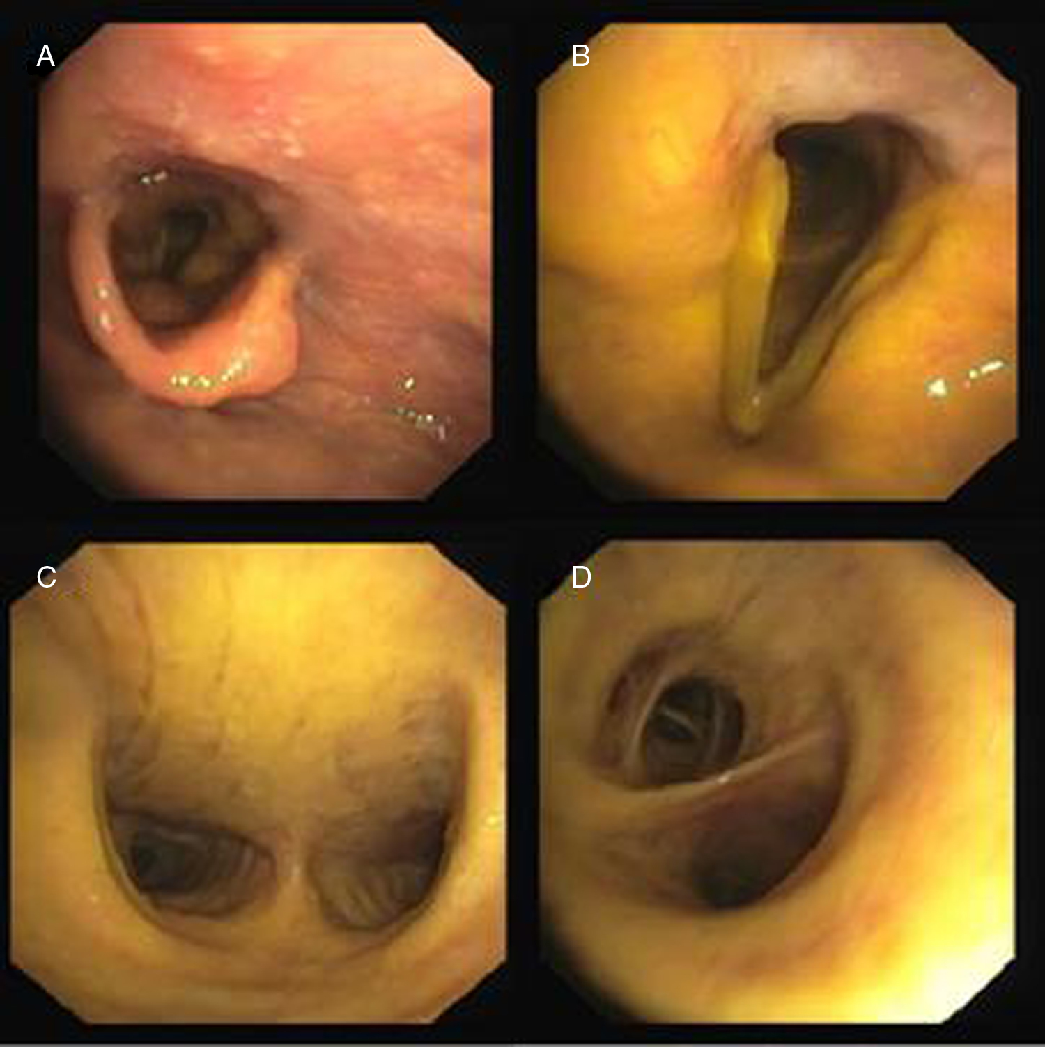
Bronchoscopy was performed during a pulmonary nodule investigation and revealed a nodule on the left side of the epiglottis and severe vocal cords and bronchial tree jaundice (Fig. 1).

Fig. 1. (A) Epiglottis; (B) Vocal cords; (C) Carina; (D) Truncus intermedius.

Bronchoscopy is not a frequently performed exam in patients with such high levels of bilirubin in the blood. In the present case bronchoscopy was performed due to the nodule location, making it difficult to access through transthoracic lung biopsy and due to the need for pulmonary metastasis exclusion in order to get a correct staging.

The high amount of elastin present in the airways makes it possible to observe a fine yellow coloration characteristic of jaundice in the bronchial tree, as observed in these endoscopic images of rare beauty and iconographic interest.

Conflict of interest

The authors have no conflicts of interest to declare.

Corresponding author. ana_f_goncalves@hotmail.com

Download PDF
Pulmonology
Article options
Tools

Are you a health professional able to prescribe or dispense drugs?