


Despite the importance of traditional pulmonary function tests (PFTs) in managing systemic sclerosis (SSc), many patients with pulmonary disease diagnosed by computed tomography (CT) present with normal PFTs.
ObjectiveTo evaluate the efficacy of the nitrogen single-breath washout (N2SBW) test in diagnosing SSc and to correlate N2SBW parameters with the PFT indexes used in the follow-up of these patients, clinical data, and CT findings.
MethodsCross-sectional study in which 52 consecutive SSc patients were subjected to spirometry, body plethysmography, analysis of the diffusing capacity for carbon monoxide (DLCO), analysis of respiratory muscle strength, N2SBW testing, and CT analysis.
ResultsTwenty-eight patients had a forced vital capacity (FVC) that was <70% of the predicted value. In the N2SBW test, 44 patients had a phase III slope (Phase III slopeN2SBW) that was >120% of the predicted value, while 15 patients had a closing volume/vital capacity (CV/VC) that was >120% of the predicted value. A significant difference in Phase III slopeN2SBW was observed when the patients with predominant traction bronchiectasis and honeycombing were compared to the patients with other CT patterns (p<0.0001). The Phase III slopeN2SBW was correlated with FVC (rs=−0.845, p<0.0001) and DLCO (rs=−0.600, p<0.0001), and the CV/VC was correlated with FVC (rs=−0.460, p=0.0006) and residual volume/total lung capacity (rs=0.328, p=0.017).
ConclusionVentilation heterogeneity is a frequent finding in SSc patients that is associated with restrictive damage, changes in pulmonary diffusion, and CT patterns. In addition, approximately one-third of the patients presented with findings that were compatible with small airway disease.
Systemic sclerosis (SSc) is a chronic inflammatory disease of the connective tissue that is characterised by cutaneous and visceral fibrosis, self-immunity, and vascular destruction.1,2 Almost 90% of SSc patients present with some form of lung injury over the evolution of the illness, and interstitial lung diseases associated with the SSc (ILD-SSc) and pulmonary arterial hypertension (PAH) are the most frequent manifestations.2,3 Among the investigation methods for ILD-SSc, lung biopsy is rarely performed. Therefore, computed tomography (CT) is currently considered the method of choice.4 Because the frequent use of ionising radiation is a matter of growing concern, CT is rarely used in the follow-up of these patients. Indeed, the severity of the pulmonary involvement of SSc is more frequently quantified using pulmonary function tests (PFTs) in clinical practice.5,6
Among the PFTs used in the diagnosis and follow-up of SSc patients, the most widespread are spirometry and diffusing capacity for carbon monoxide (DLCO).7 Despite the importance of traditional PFTs in the management of pulmonary involvement associated with SSc, a significant proportion of patients present with normal PFT results, even in the presence of ILD-SSc diagnosed by imaging methods.8 With the evolution of technical equipment in recent years, growing interest has developed in the use of the nitrogen single-breath washout (N2SBW) test to assess ventilation homogeneity and the role of small airways in several clinical conditions.9,10 The N2SBW test is used for the early diagnosis and stratification of patients and to assess the severity of several lung diseases.9,11–13 In asthma patients, poor disease control is correlated with both an increase in the closing volume (CV) and the phase III slope of the N2SBW (Phase III slopeN2SBW).13 In COPD patients, Lopes and Mafort9 observed that Phase III slopeN2SBW was the only predictor, regardless of the degree of dyspnoea and functional capacity for exercise. Mikamo et al.12 described significant correlations between the Phase III slopeN2SBW and the measurements of mechanical ventilation and emphysema score evaluated by CT. However, to the best of our knowledge, no studies have previously assessed the use of the N2SBW test in SSc patients.
In addition to causing poor ventilation distribution, lung interstitium involvement can potentially lead to structural changes in small airways, resulting in a loss of air flow that can reflect in increased ventilatory demand.14 We hypothesised that the structural disarray caused by the excessive secretion of collagen in the respiratory systems of SSc patients may be reflected in the N2SBW test. Thus, the present study sought to assess the usefulness of the N2SBW test in SSc patients and to correlate the parameters measured by the N2SBW test with the PFT indexes classically used in the follow-up of these patients, degree of dyspnoea, and CT findings.
MethodsPatientsThis was a cross-sectional study conducted between December 2015 and July 2016 in which 66 consecutive SSc patients were evaluated. These patients were recruited from the Piquet Carneiro Polyclinic of the State University of Rio de Janeiro, Rio de Janeiro, Brazil. Patients ≥18 years of age of both genders who met the criteria for SSc diagnosis15 were included in the study. The following exclusion criteria were used: patients with a previous history of smoking or those who were current smokers; individuals with asthma; evidence of overlap with other connective tissue diseases, except Sjogren's syndrome; reports of infection within the previous four weeks; and inability to perform PFTs. The protocol was approved by the Research Ethics Committee of the Pedro Ernesto University Hospital of the State University of Rio de Janeiro, Rio de Janeiro, Brazil under the number CAAE- 50752615.9.0000.5259. All of the patients signed informed consent forms.
MeasurementsDyspnoea was assessed by means of the modified Medical Research Council (mMRC) scale.16
Spirometry, body plethysmography, measurement of DLCO, and measurement of respiratory muscle strength were conducted with Collins Plus Pulmonary Function Testing Systems equipment (Warren E. Collins, Inc., Braintree, MA, USA) using the standardisation of the consensus statement.17 The Brazilian reference values were used,18–21 and the results are expressed as % predicted.
The N2SBW test was performed using the HDpft 3000 instrument (nSpire Health, Inc., Longmont, CO, USA). Briefly, individuals exhaled until the residual volume (RV) was reached and then inhaled 100% O2 until the total lung capacity was reached (TLC). Then, they slowly exhaled at a flow rate of approximately 0.3–0.5L/s until the RV was reached. The two indexes derived from the procedure are reported as % predicted22,23 and include the Phase III slopeN2SBW, which is the change in the concentration of N2 between 25% and 75% of the exhaled volume, and the closing volume/vital capacity (CV/VC), which is the portion of the VC that is exhaled after the airway begins to close. The N2SBW test was performed according to the recommendations of the consensus statement.11
We also assessed the CT scans of 31 patients that were performed within the last three months prior to recruitment. The CT scans were categorised into three patterns according to the consensus of two radiologists: grade 1 (reticular pattern predominance); grade 2 (ground-glass opacity predominance); and grade 3 (traction bronchiectasis and honeycombing predominance).4,8
Statistical analysisThe data analysis was conducted using SAS 6.11 software (SAS Institute, Inc., Cary, NC, USA). The assumption of a normal distribution of the data was evaluated with a Shapiro–Wilk test. Comparisons of variables between the two groups of patients subdivided according to the FVC or mMRC grades were evaluated by the Mann–Whitney test for numerical variables and by Fisher's exact test for categorical variables. Comparisons of the CT grades according to the different N2SBW variables were examined using the nonparametric Kruskal–Wallis test followed by Dunn's post hoc test. Spearman's rank correlation coefficient (rs) was used to evaluate the associations between the variables. The results are expressed as median values and interquartile ranges or as frequencies (percentages), and statistical significance was considered at p<0.05.
ResultsAmong the 66 patients who were considered for participation in the study, 14 were excluded for the following reasons: six for reporting a history of smoking, four for presenting with SSc together with other collagen diseases, two due to associated asthma, and two due to the inability to perform the PFTs. Thus, the study population consisted of 46 women and six men with a median age of 48 (38.5–56.3) years. Thirty-eight patients had a limited form of the disease, and 14 had the diffuse form. In 36 patients, the mMRC grade was <2 [1.12 (0.64–1.47)], and in 16, it was ≥2 [2.47 (2.31–2.78)]. In the CT analysis, the exams were categorised as grade 1 (n=13), grade 2 (n=10), and grade 3 (n=8).
For the 52 patients who participated in the study, the median values of FVC, DLCO, and FVC/DLCO were 67 (57–91)% predicted, 62 (44–81.3)% predicted, and 1.18 (0.93–1.52)% of the reference values, respectively. Twenty-eight patients had an FVC <70%, 34 had a DLCO <80%, and nine had an FVC/DLCO >1.6. Regarding the TLC, the median of the sample was 72.5 (60.3–87.5)% predicted, and this parameter was <80% in 31 patients, which indicated a restrictive disorder. The medians for Phase III slopeN2SBW and CV/VC were 254 (112–450)% predicted and 107 (62–150)% predicted, respectively. In this test, 44 patients had a Phase III slopeN2SBW >120%, while 15 patients had a CV/VC >120%. When the sample was subdivided into two groups using a cut-off value of FVC <70%, significant differences were observed between the two groups for most of the evaluated functional parameters (Table 1).
Demographic data and lung function of patients with systemic sclerosis.
| Variable | Patients with FVC ≥70% (n=24) | Patients with FVC <70% (n=28) | p-value |
|---|---|---|---|
| Demographic data | |||
| Females (%) | 22 (91.7) | 24 (85.7) | 0.67b |
| Age (years) | 49.5 (43.3–58.8) | 48 (37–53.8) | 0.26a |
| Weight (kg) | 62.8 (52.2–71) | 71.5 (57.3–82) | 0.11a |
| Height (cm) | 157 (153–162) | 160 (156–164) | 0.23a |
| BMI (kg/m2) | 25.1 (21.6–28.9) | 28.4 (22.1–30.9) | 0.15a |
| Lung function | |||
| FVC (% predicted) | 93 (80.5–103) | 57 (51.3–65) | <0.0001a |
| FEV1 (% predicted) | 87.5 (81.3–99) | 59.5 (48–65.8) | <0.0001a |
| FEV1/FVC (%) | 79.5 (74.5–84.5) | 86 (77.3–89.5) | 0.018a |
| DLCO (% predicted) | 78 (53.3–95) | 50.5 (37.3–65.8) | 0.0004a |
| FVC/DLCO (% of reference values) | 1.22 (1.08–1.52) | 1.09 (0.89–1.52) | 0.23a |
| TLC (% predicted) | 89 (79.3–98.8) | 61 (55–72) | <0.0001a |
| RV (% predicted) | 87 (73.3–109) | 74 (56.3–85) | 0.047a |
| RV/TLC (%) | 34.5 (28.2–37.2) | 40 (34.8–46.3) | 0.007a |
| Raw (cm H2O/L/s) | 1.66 (1.36–2.43) | 1.81 (1.26–3.08) | 0.78a |
| SGaw (L/s/cm H2O/L) | 0.205 (0.148–0.268) | 0.260 (0.165–0.438) | 0.12a |
| MIP (% predicted) | 66 (47–87.3) | 65 (55.3–92.8) | 0.42a |
| MEP (% predicted) | 55.5 (32.8–62) | 45.5 (34.8–54) | 0.40a |
| Nitrogen single-breath washout test | |||
| Phase III slopeN2SBW (% predicted) | 130 (88–160) | 419 (275–542) | <0.0001a |
| CV/VC (%predicted) | 71 (56–111) | 137 (101–188) | 0.003a |
Values are median (interquartile ranges) or number (%). BMI: body mass index; FVC: forced vital capacity; FEV1: forced expiratory volume in one second; DLCO: diffusing capacity for carbon monoxide; TLC: total lung capacity; RV: residual volume; Raw: airway resistance; SGaw: specific airway conductance; MIP: maximal inspiratory pressure; MEP: maximal expiratory pressure; Phase III slopeN2SBW: phase III slope of the nitrogen single-breath washout; CV/VC: closing volume/vital capacity.
The values in bold mean statistical significance.
There were no significant differences between the mMRC grades and N2SBW variables: Phase III slopeN2SBW [235 (148–287) vs. 352 (240–430), p=0.08] and CV/VC [98 (70–136) vs. 124 (85–161), p=0.13]. Regarding the CT findings, the medians of the Phase III slopeN2SBW values progressively increased from grade 1 to grade 3 [105 (64–138) vs. 185 (147–223) vs. 536 (370–653)%] with significant differences (p<0.0001). The medians of the CV/VC values also progressively increased from grade 1 to grade 3 [53 (32–85) vs. 68 (46–102) vs. 175 (78–217)%]; however, the differences were not significant (p=0.11).
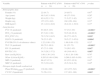
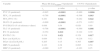
We also assessed the associations between the parameters provided by the N2SBW test, lung function indices, and CT findings (Table 2 and Figs. 1 and 2); the correlation between the Phase III slopeN2SBW and CV/VC was strong and positive (rs=0.590, p<0.0001).
Spearman's correlation coefficients for lung function parameters and nitrogen single-breath washout indexes of patients with systemic sclerosis.
| Variable | Phase III slopeN2SBW (%predicted) | CV/VC (%predicted) | ||
|---|---|---|---|---|
| rs | p-value | rs | p-value | |
| FVC (% predicted) | −0.845 | <0.0001 | −0.460 | 0.0006 |
| FEV1 (% predicted) | −0.788 | <0.0001 | −0.396 | 0.003 |
| FEV1/FVC (%) | 0.281 | 0.044 | −0.282 | 0.042 |
| DLCO (% predicted) | −0.600 | <0.0001 | −0.271 | 0.052 |
| FVC/DLCO (% of reference values) | 0.088 | 0.54 | 0.020 | 0.89 |
| TLC (% predicted) | −0.708 | <0.0001 | −0.360 | 0.008 |
| RV (% predicted) | −0.354 | 0.010 | −0.122 | 0.39 |
| RV/TLC (%) | 0.318 | 0.021 | 0.328 | 0.017 |
| Raw (cm H2O/L/s) | 0.084 | 0.55 | −0.216 | 0.12 |
| SGaw (L/s/cm H2O/L) | 0.205 | 0.15 | 0.365 | 0.007 |
| MIP (% predicted) | 0.123 | 0.38 | 0.095 | 0.50 |
| MEP (% predicted) | −0.080 | 0.57 | −0.163 | 0.25 |
Phase III slopeN2SBW: phase III slope of the nitrogen single-breath washout; CV/VC: closing volume/vital capacity; FVC: forced vital capacity; FEV1: forced expiratory volume in one second; DLCO: diffusing capacity for carbon monoxide; TLC: total lung capacity; RV: residual volume; Raw: airway resistance; SGaw: specific airway conductance; MIP: maximal inspiratory pressure; MEP: maximal expiratory pressure.
The values in bold mean statistical significance.
 and the forced vital capacity (FVC) (rs=−0.845, p<0.0001) (A) and diffusing capacity for carbon monoxide (DLCO) (rs=−0.600, p<0.0001) (B).")
 and the forced vital capacity (FVC) (rs=−0.460, p=0.0006) (A) and residual volume/total lung capacity (RV/TLC) (rs=0.328, p=0.017) (B).")
The main finding of the present study was that ventilation heterogeneity is the most common abnormality observed in the PFTs of SSc patients, and it occurs even in the absence of restrictive damage. In those patients, the more accentuated the functional or structural pulmonary deterioration is, the worse the ventilation heterogeneity. In addition, small airway disease was also a frequent finding that was related to both air trapping and the loss of lung volume. To our knowledge, this is the first study to assess the potential of the N2SBW test in SSc patients.
In the present study, the sample was almost exclusively composed of women, which is consistent with the distribution by gender reported by several investigators.2 In terms of prognosis, functional changes are important markers of the evolution of ILD-SSc, in both the initial and the sequential assessments.2 In the present study, approximately 60% of the patients had the restrictive syndrome and/or a reduction of the DLCO, thereby presenting a sample with substantial lung function involvement. Interestingly, nine patients had an FVC/DLCO >1.6, which is the recommended cut-off for study of the right side of the heart in the diagnosis of PAH.24
Many patients with pulmonary disease diagnosed by CT present with normal PFTs, which is indicative of insufficient performance of traditional PFTs in tracking pulmonary disease associated with SSc.8 In a recent study, Suliman et al.25 evaluated 102 patients with SSc and noted that 63% presented with significant ILD-SSc on CT, while only 26% had an FVC <80%. This finding emphasises the urgent need to develop new lung function parameters to diagnose lung involvement, as well as allow follow-up with patients. In this sense, some tests such as the N2SBW test, the forced oscillation technique (FOT), and impulse oscillometry can add to our understanding of the pathophysiology of ILD-SSc and can potentially be incorporated into the routine assessment of these patients.26,27
In the present study, an increase in the Phase III slopeN2SBW was the most frequent lung function abnormality, which was observed in approximately 85% of the cases and indicates the potential of this index as a marker for ILD-SSc. High values are indicative of ventilation inhomogeneity due to regional differences in time constants of the respiratory system, which result from changes in the distensibility or local resistance, thus compromising alveolar emptying.28 The increase in the Phase III slopeN2SBW in the group of patients with an FVC <70% indicates that the worsening of restrictive functional damage is an important contributor to the ventilation heterogeneity of SSc. We observed a strong association between the increase in Phase III slopeN2SBW and the decay of both the FVC and DLCO, which reinforces the routine use of these two functional indexes in the follow-up of patients with SSc in clinical practice. Interestingly, we also observed a strong association between the increase of the Phase III slopeN2SBW and the presence of traction bronchiectasis and honeycombing in CT. Despite the lack of studies correlating the N2SBW test with CT findings in fibrotic lung diseases, it is noteworthy that some researchers have observed an association between the increase in Phase III slopeN2SBW and structural lung damage in COPD patients.12,29
In addition to the Phase III slopeN2SBW, the CV/VC ratio is another index provided by the N2SBW test that has recently been studied.11,28 In the present study, we observed an increase in the CV/VC ratio in nearly one-third of the patients. A change in the CV/VC ratio has been used as one of the parameters for the diagnosis of small airway disease, and it is functionally characterised by a progressive increase in resistance as the lung is emptied, regional heterogeneity in flow rate and time constants, and premature closing of the airways.4,11 Using the FOT, Miranda et al.27 observed changes in the peripheral resistance of the respiratory system in SSc patients, which were evaluated according to the slope of the resistance as a function of frequency. Similarly, Aronsson et al.26 observed abnormalities that were compatible with small airway diseases in SSc patients including increases in the R5-R20, which is the difference between the resistance at 5Hz and the resistance at 20Hz in impulse oscillometry. Similar to the Phase III slopeN2SBW, we observed an increase in the CV/VC in the group of patients with an FVC <70%. Interestingly, we also observed a positive correlation between CV/VC and RV/TLC in the studied sample, suggesting an association between the premature closing of the airways and the presence of air trapping.30 However, it is worth emphasising that an increase in the CV/VC can be observed in patients with restrictive damage in situations where the functional residual capacity is less than the closing volume.28
It is noteworthy that there are currently more than 10 measures of ventilation distribution derived from the nitrogen washout (N2W) tests.31 The need for maintaining good coordination and cooperation when conducting VC manoeuvres under constant flow is a limiting factor for the routine use of the N2SBW test.11 Contrariwise, the multiple breath washout (MBW) test evaluates ventilation distribution during the fixed tidal volume or normal tidal breathing, assessing the release of inert gas in a series of breathing cycles. Thus, MBW shows promise for use in children and adults with difficulties performing forced manoeuvres. However, this test is time consuming, which makes it impractical to use in patients with severe lung disease.11 It is also noteworthy to highlight the double tracer gas (DTG) single-breath washout, a new N2W test modality that aims to be more specific to small airways. It distinguishes between convection-dependent and diffusion-convection-dependent ventilation heterogeneities, which occur in the conductive and acinar airways, respectively.32
The strength of this study is that it demonstrates the potential of the N2SBW test for detecting abnormalities in both ventilation and the small airways of SSc patients. Because it is a simple, non-invasive, easy and fast tool, the N2SBW test may be incorporated into the clinical assessment of SSc patients in the future. However, our study also has several limitations. First, the sample was small, and the design was cross-sectional. Second, we did not use a control group. However, the absence of a control group in this study was minimised by the use of pulmonary function values as percentages of the predicted values because these are normalised to anthropometric data. Third, the complementary use of DTG single-breath washout could have allowed us to evaluate ventilation heterogeneity in the lung periphery.32 Despite these limitations, our results provide a perspective for the use of the N2SBW test in longitudinal studies to verify its prognostic value in SSc patients.
Finally, the present study shows that in patients with SSc, ventilation distribution inhomogeneity is a very frequent finding that is related to restrictive damage, changes in pulmonary diffusion, and CT patterns. In addition, approximately one-third of the patients in this study were compatible with the criteria for small airway disease, which is associated with both the severity of restrictive damage and the presence of air trapping.
Ethical disclosuresProtection of human and animal subjectsThe authors declare that no experiments were performed on humans or animals for this study.
Confidentiality of dataThe authors declare that they have followed the protocols of their work center on the publication of patient data.
Right to privacy and informed consentThe authors have obtained the written informed consent of the patients or subjects mentioned in the article. The corresponding author is in possession of this document.
Conflicts of interestThe authors declare that they have no conflict of interest. The authors alone are responsible for the content and writing of the paper.
This research was supported by the Rio de Janeiro State Research Supporting Foundation (FAPERJ).


